 Movie
Movie Controller

[English] 日本語
 Yorodumi
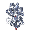
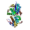
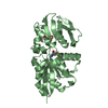
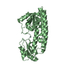
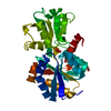
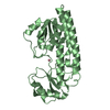
- PDB-3tmg: Crystal structure of Glycine betaine, L-proline ABC transporter, ... -
+ Open data

- Basic information
| Entry | Database: PDB / ID: 3tmg | |||||||||
|---|---|---|---|---|---|---|---|---|---|---|
| Title | Crystal structure of Glycine betaine, L-proline ABC transporter, glycine/betaine/L-proline-binding protein (ProX) from Borrelia burgdorferi | |||||||||
 Components Components | Glycine betaine, L-proline ABC transporter, glycine/betaine/L-proline-binding protein (ProX) Trimethylglycine Trimethylglycine | |||||||||
 Keywords Keywords | TRANSPORT PROTEIN / SSGCID / Structural Genomics / Seattle Structural Genomics Center for Infectious Disease | |||||||||
| Function / homology |  Function and homology information Function and homology informationtransmembrane transporter activity / ATP-binding cassette (ABC) transporter complex Similarity search - Function | |||||||||
| Biological species |  Borrelia burgdorferi (Lyme disease spirochete) Borrelia burgdorferi (Lyme disease spirochete) | |||||||||
| Method | X-RAY DIFFRACTION / SYNCHROTRON / molecular replacement / Resolution: 1.9 Å | |||||||||
 Authors Authors | Seattle Structural Genomics Center for Infectious Disease (SSGCID) | |||||||||
 Citation Citation | Journal: To be Published Title: Crystal structure of Glycine betaine, L-proline ABC transporter, glycine/betaine/L-proline-binding protein (ProX) from Borrelia burgdorferi Authors: SSGCID / Gardberg, A. / Fox, D. / Staker, B. / Stewart, L. | |||||||||
| History |
|
- Structure visualization
| Structure viewer | Molecule: MolmilJmol/JSmol |
|---|
- Downloads & links
-Download
| PDBx/mmCIF format | 3tmg.cif.gz | 437.5 KB | Display | PDBx/mmCIF format |
|---|---|---|---|---|
| PDB format | pdb3tmg.ent.gz | 358.7 KB | Display | PDB format |
| PDBx/mmJSON format | 3tmg.json.gz | Tree view | PDBx/mmJSON format | |
| Others |  Other downloads Other downloads |
-Validation report
| Arichive directory | https://data.pdbj.org/pub/pdb/validation_reports/tm/3tmgftp://data.pdbj.org/pub/pdb/validation_reports/tm/3tmg | HTTPS FTP |
|---|
-Related structure data
| Similar structure data | |
|---|---|
| Other databases |


-Links
 PDBj
PDBj

- Assembly
| Deposited unit | 
| ||||||||
|---|---|---|---|---|---|---|---|---|---|
| 1 | 
| ||||||||
| 2 | 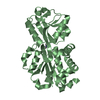
| ||||||||
| 3 | 
| ||||||||
| 4 | 
| ||||||||
| Unit cell |
|
-Components
| #1: Protein | Trimethylglycine Mass: 31786.119 Da / Num. of mol.: 4 Source method: isolated from a genetically manipulated source Source: (gene. exp.) Borrelia burgdorferi (Lyme disease spirochete)Gene: BB_0144 / Plasmid: AVA0421 / Production host: Escherichia coli (E. coli) / Strain (production host): BL21(DE3) / References: UniProt: O51169#2: Chemical | ChemComp-BET / Trimethylglycine  Mass: 118.154 Da / Num. of mol.: 4 / Source method: obtained synthetically / Formula: C5H12NO2 Mass: 118.154 Da / Num. of mol.: 4 / Source method: obtained synthetically / Formula: C5H12NO2#3: Chemical | Ethylene glycol  Mass: 62.068 Da / Num. of mol.: 2 / Source method: obtained synthetically / Formula: C2H6O2 Mass: 62.068 Da / Num. of mol.: 2 / Source method: obtained synthetically / Formula: C2H6O2#4: Chemical | ChemComp-UNL / | Num. of mol.: 1 / Source method: obtained synthetically #5: Water | ChemComp-HOH / | Water Mass: 18.015 Da / Num. of mol.: 820 / Source method: isolated from a natural source / Formula: H2O Mass: 18.015 Da / Num. of mol.: 820 / Source method: isolated from a natural source / Formula: H2O |
|---|
-Experimental details
-Experiment
| Experiment | Method: X-RAY DIFFRACTION / Number of used crystals: 1 |
|---|
- Sample preparation
| Crystal | Density Matthews: 2.38 Å3/Da / Density % sol: 48.27 % |
|---|---|
| Crystal grow | Temperature: 290 K / Method: vapor diffusion, sitting drop / pH: 6 Details: EBS internal tracking number 220361B9.PACT B9: 0.2 M LiCl, 0.1 M MES pH 6, 20% PEG 6000. BobuA.17327.a.A2 PW31644 at 26 mg/mL, vapor diffusion, sitting drop, temperature 290K, VAPOR DIFFUSION, SITTING DROP |
-Data collection
| Diffraction | Mean temperature: 100 K | ||||||||||||||||||||||||||||||||||||||||||||||||||||||||||||||||||||||||||||||||||||||||||||||||||||||||||||||||||||||||||||||||||||||||||||
|---|---|---|---|---|---|---|---|---|---|---|---|---|---|---|---|---|---|---|---|---|---|---|---|---|---|---|---|---|---|---|---|---|---|---|---|---|---|---|---|---|---|---|---|---|---|---|---|---|---|---|---|---|---|---|---|---|---|---|---|---|---|---|---|---|---|---|---|---|---|---|---|---|---|---|---|---|---|---|---|---|---|---|---|---|---|---|---|---|---|---|---|---|---|---|---|---|---|---|---|---|---|---|---|---|---|---|---|---|---|---|---|---|---|---|---|---|---|---|---|---|---|---|---|---|---|---|---|---|---|---|---|---|---|---|---|---|---|---|---|---|---|
| Diffraction source | Source: SYNCHROTRON / Site: APS  / Beamline: 21-ID-G / Wavelength: 0.97856 Å / Beamline: 21-ID-G / Wavelength: 0.97856 Å | ||||||||||||||||||||||||||||||||||||||||||||||||||||||||||||||||||||||||||||||||||||||||||||||||||||||||||||||||||||||||||||||||||||||||||||
| Detector | Type: MARMOSAIC 300 mm CCD / Detector: CCD / Date: Mar 7, 2011 | ||||||||||||||||||||||||||||||||||||||||||||||||||||||||||||||||||||||||||||||||||||||||||||||||||||||||||||||||||||||||||||||||||||||||||||
| Radiation | Monochromator: double crystal / Protocol: SINGLE WAVELENGTH / Monochromatic (M) / Laue (L): M / Scattering type: x-ray | ||||||||||||||||||||||||||||||||||||||||||||||||||||||||||||||||||||||||||||||||||||||||||||||||||||||||||||||||||||||||||||||||||||||||||||
| Radiation wavelength | Wavelength: 0.97856 Å / Relative weight: 1 | ||||||||||||||||||||||||||||||||||||||||||||||||||||||||||||||||||||||||||||||||||||||||||||||||||||||||||||||||||||||||||||||||||||||||||||
| Reflection | Resolution: 1.9→49.16 Å / Num. all: 91173 / Num. obs: 88358 / % possible obs: 96.9 % / Observed criterion σ(F): 0 / Observed criterion σ(I): -3 / Redundancy: 3.9 % / Biso Wilson estimate: 26.791 Å2 / Rmerge(I) obs: 0.086 / Net I/σ(I): 17.74 | ||||||||||||||||||||||||||||||||||||||||||||||||||||||||||||||||||||||||||||||||||||||||||||||||||||||||||||||||||||||||||||||||||||||||||||
| Reflection shell | Diffraction-ID: 1
|
-Phasing
| Phasing | Method: molecular replacement | ||||||
|---|---|---|---|---|---|---|---|
| Phasing MR | Model details: Phaser MODE: MR_AUTO
|
- Processing
| Software |
| |||||||||||||||||||||||||||||||||||||||||||||||||||||||||||||||||||||||||||||||||||||||||||||||||||||||||||||||||||||||||||||
|---|---|---|---|---|---|---|---|---|---|---|---|---|---|---|---|---|---|---|---|---|---|---|---|---|---|---|---|---|---|---|---|---|---|---|---|---|---|---|---|---|---|---|---|---|---|---|---|---|---|---|---|---|---|---|---|---|---|---|---|---|---|---|---|---|---|---|---|---|---|---|---|---|---|---|---|---|---|---|---|---|---|---|---|---|---|---|---|---|---|---|---|---|---|---|---|---|---|---|---|---|---|---|---|---|---|---|---|---|---|---|---|---|---|---|---|---|---|---|---|---|---|---|---|---|---|---|
| Refinement | Method to determine structure: molecular replacement / Resolution: 1.9→49.16 Å / Cor.coef. Fo:Fc: 0.955 / Cor.coef. Fo:Fc free: 0.93 / SU B: 5.83 / SU ML: 0.09 / Cross valid method: THROUGHOUT / σ(F): 0 / ESU R Free: 0.139 / Stereochemistry target values: MAXIMUM LIKELIHOOD Details: U VALUES: WITH TLS ADDED HYDROGENS HAVE BEEN USED IF PRESENT IN THE INPUT
| |||||||||||||||||||||||||||||||||||||||||||||||||||||||||||||||||||||||||||||||||||||||||||||||||||||||||||||||||||||||||||||
| Solvent computation | Ion probe radii: 0.8 Å / Shrinkage radii: 0.8 Å / VDW probe radii: 1.2 Å / Solvent model: MASK | |||||||||||||||||||||||||||||||||||||||||||||||||||||||||||||||||||||||||||||||||||||||||||||||||||||||||||||||||||||||||||||
| Displacement parameters | Biso mean: 22.682 Å2
| |||||||||||||||||||||||||||||||||||||||||||||||||||||||||||||||||||||||||||||||||||||||||||||||||||||||||||||||||||||||||||||
| Refinement step | Cycle: LAST / Resolution: 1.9→49.16 Å
| |||||||||||||||||||||||||||||||||||||||||||||||||||||||||||||||||||||||||||||||||||||||||||||||||||||||||||||||||||||||||||||
| Refine LS restraints |
| |||||||||||||||||||||||||||||||||||||||||||||||||||||||||||||||||||||||||||||||||||||||||||||||||||||||||||||||||||||||||||||
| LS refinement shell | Resolution: 1.9→1.949 Å / Total num. of bins used: 20
| |||||||||||||||||||||||||||||||||||||||||||||||||||||||||||||||||||||||||||||||||||||||||||||||||||||||||||||||||||||||||||||
| Refinement TLS params. | Method: refined / Refine-ID: X-RAY DIFFRACTION
| |||||||||||||||||||||||||||||||||||||||||||||||||||||||||||||||||||||||||||||||||||||||||||||||||||||||||||||||||||||||||||||
| Refinement TLS group |
|
