 Movie
Movie Controller

[English] 日本語
 Yorodumi
- PDB-3tm6: Crystal structure of the beta-2 microglobulin DIMC50 disulphide-l... -
+ Open data

- Basic information
| Entry | Database: PDB / ID: 3tm6 | ||||||
|---|---|---|---|---|---|---|---|
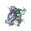
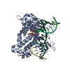
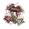
| Title | Crystal structure of the beta-2 microglobulin DIMC50 disulphide-linked homodimer mutant | ||||||
 Components Components | Beta-2-microglobulin Beta-2 microglobulin Beta-2 microglobulin | ||||||
 Keywords Keywords | IMMUNE SYSTEM / Immunoglobulin-like fold / MHC class I / Light chain | ||||||
| Function / homology |  Function and homology information Function and homology informationpositive regulation of ferrous iron binding / positive regulation of transferrin receptor binding / positive regulation of receptor binding / early endosome lumen / Nef mediated downregulation of MHC class I complex cell surface expression / DAP12 interactions / negative regulation of receptor binding / Endosomal/Vacuolar pathway / Antigen Presentation: Folding, assembly and peptide loading of class I MHC / cellular response to iron(III) ion ...positive regulation of ferrous iron binding / positive regulation of transferrin receptor binding / positive regulation of receptor binding / early endosome lumen / Nef mediated downregulation of MHC class I complex cell surface expression / DAP12 interactions / negative regulation of receptor binding / Endosomal/Vacuolar pathway / Antigen Presentation: Folding, assembly and peptide loading of class I MHC / cellular response to iron(III) ion / antigen processing and presentation of exogenous protein antigen via MHC class Ib, TAP-dependent / negative regulation of forebrain neuron differentiation / ER to Golgi transport vesicle membrane / regulation of erythrocyte differentiation / peptide antigen assembly with MHC class I protein complex / response to molecule of bacterial origin / regulation of iron ion transport / MHC class I peptide loading complex / HFE-transferrin receptor complex / T cell mediated cytotoxicity / cellular response to iron ion / antigen processing and presentation of endogenous peptide antigen via MHC class I / positive regulation of T cell cytokine production / MHC class I protein complex / multicellular organismal-level iron ion homeostasis / negative regulation of neurogenesis / peptide antigen assembly with MHC class II protein complex / positive regulation of receptor-mediated endocytosis / positive regulation of T cell mediated cytotoxicity / MHC class II protein complex / cellular response to nicotine / recycling endosome membrane / specific granule lumen / phagocytic vesicle membrane / peptide antigen binding / positive regulation of cellular senescence / antigen processing and presentation of exogenous peptide antigen via MHC class II / negative regulation of epithelial cell proliferation / Immunoregulatory interactions between a Lymphoid and a non-Lymphoid cell / Interferon gamma signaling / positive regulation of immune response / Modulation by Mtb of host immune system / sensory perception of smell / positive regulation of T cell activation / positive regulation of protein binding / tertiary granule lumen / DAP12 signaling / negative regulation of neuron projection development / MHC class II protein complex binding / late endosome membrane / T cell differentiation in thymus / ER-Phagosome pathway / iron ion transport / early endosome membrane / protein refolding / protein homotetramerization / intracellular iron ion homeostasis / amyloid fibril formation / learning or memory / Amyloid fiber formation / lysosomal membrane / external side of plasma membrane / endoplasmic reticulum lumen / Golgi membrane / focal adhesion / Neutrophil degranulation / SARS-CoV-2 activates/modulates innate and adaptive immune responses / structural molecule activity / Golgi apparatus / endoplasmic reticulum / protein homodimerization activity / extracellular space / extracellular exosome / extracellular region / membrane / identical protein binding / plasma membrane / cytosolSimilarity search - Function | ||||||
| Biological species |  Homo sapiens (human) Homo sapiens (human) | ||||||
| Method | X-RAY DIFFRACTION / SYNCHROTRON / MOLECULAR REPLACEMENT / Resolution: 2.7 Å | ||||||
 Authors Authors | Colombo, M. / Ricagno, S. / Bolognesi, M. | ||||||
 Citation Citation | Journal: Febs J. / Year: 2012 Title: A recurrent D-strand association interface is observed in beta-2 microglobulin oligomers. Authors: Colombo, M. / de Rosa, M. / Bellotti, V. / Ricagno, S. / Bolognesi, M. | ||||||
| History |
|
- Structure visualization
| Structure viewer | Molecule: MolmilJmol/JSmol |
|---|
- Downloads & links
-Download
| PDBx/mmCIF format | 3tm6.cif.gz | 173.1 KB | Display | PDBx/mmCIF format |
|---|---|---|---|---|
| PDB format | pdb3tm6.ent.gz | 139.1 KB | Display | PDB format |
| PDBx/mmJSON format | 3tm6.json.gz | Tree view | PDBx/mmJSON format | |
| Others |  Other downloads Other downloads |
-Validation report
| Arichive directory | https://data.pdbj.org/pub/pdb/validation_reports/tm/3tm6ftp://data.pdbj.org/pub/pdb/validation_reports/tm/3tm6 | HTTPS FTP |
|---|
-Related structure data
| Related structure data |  3tlrC 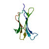 2z9tS S: Starting model for refinement C: citing same article ( |
|---|---|
| Similar structure data |


-Links
 PDBj
PDBj

- Assembly
| Deposited unit | 
| ||||||||
|---|---|---|---|---|---|---|---|---|---|
| 1 | 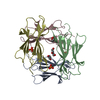
| ||||||||
| 2 | 
| ||||||||
| Unit cell |
|
-Components
| #1: Protein | Beta-2 microglobulin / Beta-2-microglobulin form pI 5.3 Mass: 11853.386 Da / Num. of mol.: 8 / Fragment: Full-length Beta-2 microglobulin / Mutation: E50C Source method: isolated from a genetically manipulated source Source: (gene. exp.) Homo sapiens (human) / Gene: B2M, CDABP0092, HDCMA22P / Production host:  Escherichia coli (E. coli) / References: UniProt: P61769 Escherichia coli (E. coli) / References: UniProt: P61769#2: Chemical | ChemComp-PEG / Diethylene glycol  Mass: 106.120 Da / Num. of mol.: 5 / Source method: obtained synthetically / Formula: C4H10O3 Mass: 106.120 Da / Num. of mol.: 5 / Source method: obtained synthetically / Formula: C4H10O3#3: Chemical | ChemComp-PO4 / Phosphate  Mass: 94.971 Da / Num. of mol.: 6 / Source method: obtained synthetically / Formula: PO4 Mass: 94.971 Da / Num. of mol.: 6 / Source method: obtained synthetically / Formula: PO4#4: Water | ChemComp-HOH / | Water Mass: 18.015 Da / Num. of mol.: 112 / Source method: isolated from a natural source / Formula: H2O Mass: 18.015 Da / Num. of mol.: 112 / Source method: isolated from a natural source / Formula: H2O |
|---|
-Experimental details
-Experiment
| Experiment | Method: X-RAY DIFFRACTION / Number of used crystals: 1 |
|---|
- Sample preparation
| Crystal | Density Matthews: 2.32 Å3/Da / Density % sol: 47.03 % |
|---|---|
| Crystal grow | Temperature: 293 K / Method: vapor diffusion, sitting drop / pH: 5.5 Details: imidazole-malate 0.2 M, pH 5.5, PEG 600 24%, VAPOR DIFFUSION, SITTING DROP, temperature 293K |
-Data collection
| Diffraction | Mean temperature: 100 K |
|---|---|
| Diffraction source | Source: SYNCHROTRON / Site: ESRF  / Beamline: ID14-4 / Wavelength: 0.9395 Å / Beamline: ID14-4 / Wavelength: 0.9395 Å |
| Detector | Type: ADSC QUANTUM 315r / Detector: CCD / Date: Nov 7, 2009 |
| Radiation | Monochromator: channel cut ESRF monochromator / Protocol: SINGLE WAVELENGTH / Monochromatic (M) / Laue (L): M / Scattering type: x-ray |
| Radiation wavelength | Wavelength: 0.9395 Å / Relative weight: 1 |
| Reflection | Resolution: 2.7→29.39 Å / Num. all: 24109 / Num. obs: 23049 / % possible obs: 95.6 % / Observed criterion σ(I): -3 / Biso Wilson estimate: 65.69 Å2 |
| Reflection shell | Resolution: 2.7→2.85 Å / % possible all: 97.4 |
- Processing
| Software |
| ||||||||||||||||||||||||||||||||||||||||||||||||||||||||||||||||||
|---|---|---|---|---|---|---|---|---|---|---|---|---|---|---|---|---|---|---|---|---|---|---|---|---|---|---|---|---|---|---|---|---|---|---|---|---|---|---|---|---|---|---|---|---|---|---|---|---|---|---|---|---|---|---|---|---|---|---|---|---|---|---|---|---|---|---|---|
| Refinement | Method to determine structure: MOLECULAR REPLACEMENT Starting model: 2Z9T Resolution: 2.7→29.39 Å / Cor.coef. Fo:Fc: 0.8738 / Cor.coef. Fo:Fc free: 0.8379 / Cross valid method: THROUGHOUT / σ(F): 0 / Stereochemistry target values: Engh & Huber
| ||||||||||||||||||||||||||||||||||||||||||||||||||||||||||||||||||
| Displacement parameters | Biso mean: 43.95 Å2
| ||||||||||||||||||||||||||||||||||||||||||||||||||||||||||||||||||
| Refine analyze | Luzzati coordinate error obs: 0.419 Å | ||||||||||||||||||||||||||||||||||||||||||||||||||||||||||||||||||
| Refinement step | Cycle: LAST / Resolution: 2.7→29.39 Å
| ||||||||||||||||||||||||||||||||||||||||||||||||||||||||||||||||||
| Refine LS restraints |
| ||||||||||||||||||||||||||||||||||||||||||||||||||||||||||||||||||
| LS refinement shell | Resolution: 2.7→2.82 Å / Total num. of bins used: 12
|
