 Movie
Movie Controller

[English] 日本語
 Yorodumi
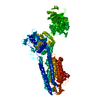
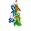
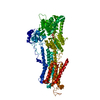
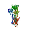
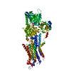
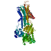
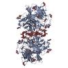
- PDB-3tlm: Crystal Structure of Endoplasmic Reticulum Ca2+-ATPase (SERCA) Fr... -
+ Open data

- Basic information
| Entry | Database: PDB / ID: 3tlm | ||||||
|---|---|---|---|---|---|---|---|
| Title | Crystal Structure of Endoplasmic Reticulum Ca2+-ATPase (SERCA) From Bovine Muscle | ||||||
 Components Components | Sarcoplasmic/endoplasmic reticulum calcium ATPase 1 | ||||||
 Keywords Keywords |  HYDROLASE / SERCA / Ca-ATPase / Calcium transporter / Ca / Fast-twitch muscle HYDROLASE / SERCA / Ca-ATPase / Calcium transporter / Ca / Fast-twitch muscle | ||||||
| Function / homology |  Function and homology information Function and homology informationmaintenance of mitochondrion location / relaxation of skeletal muscle / positive regulation of endoplasmic reticulum calcium ion concentration / positive regulation of mitochondrial calcium ion concentration / positive regulation of cardiac muscle cell contraction / positive regulation of calcium ion import into sarcoplasmic reticulum / positive regulation of fast-twitch skeletal muscle fiber contraction / P-type proton-exporting transporter activity / calcium ion import into sarcoplasmic reticulum / negative regulation of endoplasmic reticulum calcium ion concentration ...maintenance of mitochondrion location / relaxation of skeletal muscle / positive regulation of endoplasmic reticulum calcium ion concentration / positive regulation of mitochondrial calcium ion concentration / positive regulation of cardiac muscle cell contraction / positive regulation of calcium ion import into sarcoplasmic reticulum / positive regulation of fast-twitch skeletal muscle fiber contraction / P-type proton-exporting transporter activity / calcium ion import into sarcoplasmic reticulum / negative regulation of endoplasmic reticulum calcium ion concentration / positive regulation of ATPase-coupled calcium transmembrane transporter activity / P-type Ca2+ transporter / P-type calcium transporter activity / apoptotic mitochondrial changes / intrinsic apoptotic signaling pathway in response to endoplasmic reticulum stress / sarcoplasmic reticulum membrane / calcium ion transmembrane transport / intracellular calcium ion homeostasis / membrane => GO:0016020 / calcium ion binding / ATP hydrolysis activity / protein homodimerization activity / mitochondrion / ATP bindingSimilarity search - Function | ||||||
| Biological species |  Bos taurus (cattle) Bos taurus (cattle) | ||||||
| Method | X-RAY DIFFRACTION / SYNCHROTRON / MOLECULAR REPLACEMENT / Resolution: 2.95 Å | ||||||
 Authors Authors | Sacchetto, R. / Bertipaglia, I. / Giannetti, S. / Cendron, L. / Mascarello, F. / Damiani, E. / Carafoli, E. / Zanotti, G. | ||||||
 Citation Citation | Journal: J.Struct.Biol. / Year: 2012 Title: Crystal structure of sarcoplasmic reticulum Ca(2+)-ATPase (SERCA) from bovine muscle. Authors: Sacchetto, R. / Bertipaglia, I. / Giannetti, S. / Cendron, L. / Mascarello, F. / Damiani, E. / Carafoli, E. / Zanotti, G. #1: Journal: Nature / Year: 2000Title: Crystal structure of the calcium pump of sarcoplasmic reticulum at 2.6 A resolution. Authors: Toyoshima, C. / Nakasako, M. / Nomura, H. / Ogawa, H. #2: Journal: Nature / Year: 2007Title: The structural basis of calcium transport by the calcium pump. Authors: Olesen, C. / Picard, M. / Winther, A.M. / Gyrup, C. / Morth, J.P. / Oxvig, C. / Moller, J.V. / Nissen, P. #3: Journal: Physiol Rev / Year: 2009 Title: Calcium pumps in health and disease. Authors: Brini, M. / Carafoli, E. | ||||||
| History |
|
- Structure visualization
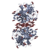
| Structure viewer | Molecule: MolmilJmol/JSmol |
|---|
- Downloads & links
-Download
| PDBx/mmCIF format | 3tlm.cif.gz | 209 KB | Display | PDBx/mmCIF format |
|---|---|---|---|---|
| PDB format | pdb3tlm.ent.gz | 162.6 KB | Display | PDB format |
| PDBx/mmJSON format | 3tlm.json.gz | Tree view | PDBx/mmJSON format | |
| Others |  Other downloads Other downloads |
-Validation report
| Arichive directory | https://data.pdbj.org/pub/pdb/validation_reports/tl/3tlmftp://data.pdbj.org/pub/pdb/validation_reports/tl/3tlm | HTTPS FTP |
|---|
-Related structure data
| Related structure data | 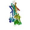 1t5sS S: Starting model for refinement |
|---|---|
| Similar structure data |

-Links
 PDBj
PDBj


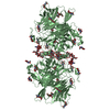
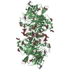
- Assembly
| Deposited unit | 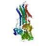
| ||||||||
|---|---|---|---|---|---|---|---|---|---|
| 1 |
| ||||||||
| Unit cell |
|
-Components
-Protein , 1 types, 1 molecules A
| #1: Protein | Mass: 109346.297 Da / Num. of mol.: 1 / Source method: isolated from a natural source / Details: cutaneus trunci muscle / Source: (natural) Bos taurus (cattle) / Strain: Chianina cattle / References: UniProt: Q0VCY0, EC: 3.6.3.8 |
|---|
-Non-polymers , 5 types, 38 molecules 


| #2: Chemical |  Mass: 40.078 Da / Num. of mol.: 2 / Source method: obtained synthetically / Formula: Ca Mass: 40.078 Da / Num. of mol.: 2 / Source method: obtained synthetically / Formula: Ca#3: Chemical | ChemComp-MG / |  Mass: 24.305 Da / Num. of mol.: 1 / Source method: obtained synthetically / Formula: Mg Mass: 24.305 Da / Num. of mol.: 1 / Source method: obtained synthetically / Formula: Mg#4: Chemical | ChemComp-K / |  Mass: 39.098 Da / Num. of mol.: 1 / Source method: obtained synthetically / Formula: K Mass: 39.098 Da / Num. of mol.: 1 / Source method: obtained synthetically / Formula: K#5: Chemical | ChemComp-ACP / |  Mass: 505.208 Da / Num. of mol.: 1 / Source method: obtained synthetically / Formula: C11H18N5O12P3 / Comment: AMP-PCP, energy-carrying molecule analogue*YM Mass: 505.208 Da / Num. of mol.: 1 / Source method: obtained synthetically / Formula: C11H18N5O12P3 / Comment: AMP-PCP, energy-carrying molecule analogue*YM#6: Water | ChemComp-HOH / | WaterMass: 18.015 Da / Num. of mol.: 33 / Source method: isolated from a natural source / Formula: H2O |
|---|
-Experimental details
-Experiment
| Experiment | Method: X-RAY DIFFRACTION / Number of used crystals: 2 |
|---|
- Sample preparation
| Crystal | Density Matthews: 3.88 Å3/Da / Density % sol: 68.32 % |
|---|---|
| Crystal grow | Temperature: 292 K / Method: vapor diffusion, hanging drop Details: 220mM sodium acetate, 4% t-butanol, 5mM mercaptoethanol, 15% glycerol, 6-7% PEG 6000, VAPOR DIFFUSION, HANGING DROP, temperature 292K |
-Data collection
| Diffraction | Mean temperature: 100 K |
|---|---|
| Diffraction source | Source: SYNCHROTRON / Site: ESRF  / Beamline: ID29 / Wavelength: 1.1271 Å / Beamline: ID29 / Wavelength: 1.1271 Å |
| Detector | Type: ADSC QUANTUM 315r / Detector: CCD / Date: Oct 29, 2009 |
| Radiation | Protocol: SINGLE WAVELENGTH / Monochromatic (M) / Laue (L): M / Scattering type: x-ray |
| Radiation wavelength | Wavelength: 1.1271 Å / Relative weight: 1 |
| Reflection | Resolution: 2.95→76.61 Å / Num. all: 35944 / Num. obs: 35546 / % possible obs: 99.1 % / Observed criterion σ(F): 0 / Observed criterion σ(I): 0 / Redundancy: 7.4 % / Biso Wilson estimate: 60.2 Å2 / Rmerge(I) obs: 0.106 / Net I/σ(I): 14.1 |
| Reflection shell | Resolution: 2.95→3.11 Å / Redundancy: 7.5 % / Rmerge(I) obs: 0.692 / Mean I/σ(I) obs: 3.5 / % possible all: 98.4 |
- Processing
| Software |
| ||||||||||||||||||||||||||||||||||||||||||||||||||||||||||||
|---|---|---|---|---|---|---|---|---|---|---|---|---|---|---|---|---|---|---|---|---|---|---|---|---|---|---|---|---|---|---|---|---|---|---|---|---|---|---|---|---|---|---|---|---|---|---|---|---|---|---|---|---|---|---|---|---|---|---|---|---|---|
| Refinement | Method to determine structure: MOLECULAR REPLACEMENT Starting model: PDB ENTRY 1T5S Resolution: 2.95→41.99 Å / Rfactor Rfree error: 0.007 / Data cutoff high absF: 2964064.54 / Data cutoff low absF: 0 / Isotropic thermal model: RESTRAINED / Cross valid method: THROUGHOUT / σ(F): 0 / Stereochemistry target values: Engh & Huber / Details: BULK SOLVENT MODEL USED
| ||||||||||||||||||||||||||||||||||||||||||||||||||||||||||||
| Solvent computation | Solvent model: FLAT MODEL / Bsol: 44.0516 Å2 / ksol: 0.3 e/Å3 | ||||||||||||||||||||||||||||||||||||||||||||||||||||||||||||
| Displacement parameters | Biso mean: 80.2 Å2
| ||||||||||||||||||||||||||||||||||||||||||||||||||||||||||||
| Refine analyze |
| ||||||||||||||||||||||||||||||||||||||||||||||||||||||||||||
| Refinement step | Cycle: LAST / Resolution: 2.95→41.99 Å
| ||||||||||||||||||||||||||||||||||||||||||||||||||||||||||||
| Refine LS restraints |
| ||||||||||||||||||||||||||||||||||||||||||||||||||||||||||||
| LS refinement shell | Resolution: 2.95→3.13 Å / Rfactor Rfree error: 0.02 / Total num. of bins used: 6
| ||||||||||||||||||||||||||||||||||||||||||||||||||||||||||||
| Xplor file |
|
