 Movie
Movie Controller

[English] 日本語
 Yorodumi
- PDB-3tk3: Cytochrome P450 2B4 mutant L437A in complex with 4-(4-chloropheny... -
+ Open data

- Basic information
| Entry | Database: PDB / ID: 3tk3 | ||||||
|---|---|---|---|---|---|---|---|
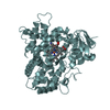
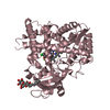
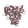
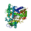
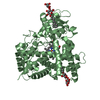
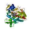
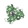
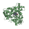
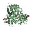
| Title | Cytochrome P450 2B4 mutant L437A in complex with 4-(4-chlorophenyl)imidazole | ||||||
 Components Components | Cytochrome P450 2B4 | ||||||
 Keywords Keywords |  OXIDOREDUCTASE / P450 / cytochrome P450 2B4 / monooxygenase / membrane protein / CYP 2B4 / CYP LM2 OXIDOREDUCTASE / P450 / cytochrome P450 2B4 / monooxygenase / membrane protein / CYP 2B4 / CYP LM2 | ||||||
| Function / homology |  Function and homology information Function and homology informationresponse to stimulus / unspecific monooxygenase / aromatase activity / iron ion binding / heme binding / endoplasmic reticulum membraneSimilarity search - Function | ||||||
| Biological species |  Oryctolagus cuniculus (rabbit) Oryctolagus cuniculus (rabbit) | ||||||
| Method | X-RAY DIFFRACTION / SYNCHROTRON / MOLECULAR REPLACEMENT / molecular replacement / Resolution: 2.8001 Å | ||||||
 Authors Authors | Gay, S.C. / Jang, H.H. / Wilderman, P.R. / Zhang, Q. / Stout, C.D. / Halpert, J.R. | ||||||
 Citation Citation | Journal: Febs J. / Year: 2012 Title: Investigation by site-directed mutagenesis of the role of cytochrome P450 2B4 non-active-site residues in protein-ligand interactions based on crystal structures of the ligand-bound enzyme. Authors: Wilderman, P.R. / Gay, S.C. / Jang, H.H. / Zhang, Q. / Stout, C.D. / Halpert, J.R. #1: Journal: J.Biol.Chem. / Year: 2006Title: Structure of microsomal cytochrome P450 2B4 complexed with the antifungal drug bifonazole: insight into P450 conformational plasticity and membrane interaction. Authors: Zhao, Y. / White, M.A. / Muralidhara, B.K. / Sun, L. / Halpert, J.R. / Stout, C.D. #2: Journal: J.Biol.Chem. / Year: 2004Title: Structure of mammalian cytochrome P450 2B4 complexed with 4-(4-chlorophenyl)imidazole at 1.9-A resolution: insight into the range of P450 conformations and the coordination of redox partner binding. Authors: Scott, E.E. / White, M.A. / He, Y.A. / Johnson, E.F. / Stout, C.D. / Halpert, J.R. #3: Journal: Proc.Natl.Acad.Sci.USA / Year: 2003Title: An open conformation of mammalian cytochrome P450 2B4 at 1.6-A resolution. Authors: Scott, E.E. / He, Y.A. / Wester, M.R. / White, M.A. / Chin, C.C. / Halpert, J.R. / Johnson, E.F. / Stout, C.D. #4: Journal: Biochemistry / Year: 2007Title: Structural and thermodynamic consequences of 1-(4-chlorophenyl)imidazole binding to cytochrome P450 2B4. Authors: Zhao, Y. / Sun, L. / Muralidhara, B.K. / Kumar, S. / White, M.A. / Stout, C.D. / Halpert, J.R. #5: Journal: Biochemistry / Year: 2009Title: Crystal structures of cytochrome P450 2B4 in complex with the inhibitor 1-biphenyl-4-methyl-1H-imidazole: ligand-induced structural response through alpha-helical repositioning. Authors: Gay, S.C. / Sun, L. / Maekawa, K. / Halpert, J.R. / Stout, C.D. #6: Journal: Biochemistry / Year: 2010Title: Structures of cytochrome P450 2B4 complexed with the antiplatelet drugs ticlopidine and clopidogrel . Authors: Gay, S.C. / Roberts, A.G. / Maekawa, K. / Talakad, J.C. / Hong, W.X. / Zhang, Q. / Stout, C.D. / Halpert, J.R. #7: Journal: J.Biol.Chem. / Year: 2010Title: Plasticity of cytochrome P450 2B4 as investigated by hydrogen-deuterium exchange mass spectrometry and X-ray crystallography. Authors: Wilderman, P.R. / Shah, M.B. / Liu, T. / Li, S. / Hsu, S. / Roberts, A.G. / Goodlett, D.R. / Zhang, Q. / Woods, V.L. / Stout, C.D. / Halpert, J.R. #8: Journal: Biochemistry / Year: 2011Title: Structural Analysis of Mammalian Cytochrome P450 2B4 Covalently Bound to the Mechanism-Based Inactivator tert-Butylphenylacetylene: Insight into Partial Enzymatic Activity Authors: C Gay, S. / Zhang, H. / Wilderman, P.R. / Roberts, A.G. / Liu, T. / Li, S. / Lin, H.L. / Woods Jr., V.L. / Stout, C.D. / Hollenberg, P.F. / Halpert, J.R. | ||||||
| History |
|
- Structure visualization
| Structure viewer | Molecule: MolmilJmol/JSmol |
|---|
- Downloads & links
-Download
| PDBx/mmCIF format | 3tk3.cif.gz | 383.6 KB | Display | PDBx/mmCIF format |
|---|---|---|---|---|
| PDB format | pdb3tk3.ent.gz | 311.8 KB | Display | PDB format |
| PDBx/mmJSON format | 3tk3.json.gz | Tree view | PDBx/mmJSON format | |
| Others |  Other downloads Other downloads |
-Validation report
| Arichive directory | https://data.pdbj.org/pub/pdb/validation_reports/tk/3tk3ftp://data.pdbj.org/pub/pdb/validation_reports/tk/3tk3 | HTTPS FTP |
|---|
-Related structure data
| Related structure data | 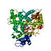 1suoS S: Starting model for refinement |
|---|---|
| Similar structure data |

-Links
 PDBj
PDBj


- Assembly
| Deposited unit | 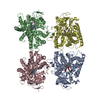
| ||||||||
|---|---|---|---|---|---|---|---|---|---|
| 1 | 
| ||||||||
| 2 | 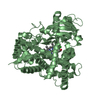
| ||||||||
| 3 | 
| ||||||||
| 4 | 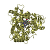
| ||||||||
| Unit cell |
|
-Components
| #1: Protein | Mass: 54127.004 Da / Num. of mol.: 4 Mutation: E2A, G22K, H23K, P24T, K25S, A26S, H27K, R29K, H226Y, L437A Source method: isolated from a genetically manipulated source Source: (gene. exp.) Oryctolagus cuniculus (rabbit) / Gene: CYP2B4 / Plasmid: PKK / Production host:  Escherichia coli (E. coli) / Strain (production host): TOPP3 / References: UniProt: P00178, unspecific monooxygenase Escherichia coli (E. coli) / Strain (production host): TOPP3 / References: UniProt: P00178, unspecific monooxygenase#2: Chemical | ChemComp-HEM / Heme B  Mass: 616.487 Da / Num. of mol.: 4 / Source method: obtained synthetically / Formula: C34H32FeN4O4 Mass: 616.487 Da / Num. of mol.: 4 / Source method: obtained synthetically / Formula: C34H32FeN4O4#3: Chemical | ChemComp-CPZ /   Mass: 178.618 Da / Num. of mol.: 4 / Source method: obtained synthetically / Formula: C9H7ClN2 Mass: 178.618 Da / Num. of mol.: 4 / Source method: obtained synthetically / Formula: C9H7ClN2#4: Water | ChemComp-HOH / | Water Mass: 18.015 Da / Num. of mol.: 290 / Source method: isolated from a natural source / Formula: H2O Mass: 18.015 Da / Num. of mol.: 290 / Source method: isolated from a natural source / Formula: H2OSequence details | AUTHORS INFORMED THAT THE GENBANK SEQUENCE IS THOUGHT TO CONTAIN A SEQUENCING | |
|---|
-Experimental details
-Experiment
| Experiment | Method: X-RAY DIFFRACTION / Number of used crystals: 1 |
|---|
- Sample preparation
| Crystal | Density Matthews: 4.12 Å3/Da / Density % sol: 70.14 % |
|---|---|
| Crystal grow | Temperature: 291 K / Method: vapor diffusion, sitting drop / pH: 6 Details: 0.1 M MES, 20% (w/v) PEG 8000, 0.2 M calcium acetate, pH 6.0, vapor diffusion, sitting drop, temperature 291K |
-Data collection
| Diffraction | Mean temperature: 100 K | ||||||||||||||||||||||||||||||||||||||||||||||||||||||||||||||||||||||||||||||||||||||||
|---|---|---|---|---|---|---|---|---|---|---|---|---|---|---|---|---|---|---|---|---|---|---|---|---|---|---|---|---|---|---|---|---|---|---|---|---|---|---|---|---|---|---|---|---|---|---|---|---|---|---|---|---|---|---|---|---|---|---|---|---|---|---|---|---|---|---|---|---|---|---|---|---|---|---|---|---|---|---|---|---|---|---|---|---|---|---|---|---|---|
| Diffraction source | Source: SYNCHROTRON / Site: SSRL  / Beamline: BL11-1 / Wavelength: 0.98 Å / Beamline: BL11-1 / Wavelength: 0.98 Å | ||||||||||||||||||||||||||||||||||||||||||||||||||||||||||||||||||||||||||||||||||||||||
| Detector | Type: MARMOSAIC 325 mm CCD / Detector: CCD / Date: Jul 2, 2011 | ||||||||||||||||||||||||||||||||||||||||||||||||||||||||||||||||||||||||||||||||||||||||
| Radiation | Monochromator: Si(111) / Protocol: SINGLE WAVELENGTH / Monochromatic (M) / Laue (L): M / Scattering type: x-ray | ||||||||||||||||||||||||||||||||||||||||||||||||||||||||||||||||||||||||||||||||||||||||
| Radiation wavelength | Wavelength: 0.98 Å / Relative weight: 1 | ||||||||||||||||||||||||||||||||||||||||||||||||||||||||||||||||||||||||||||||||||||||||
| Reflection | Resolution: 2.8→76.24 Å / Num. all: 85144 / Num. obs: 84142 / % possible obs: 99.4 % / Observed criterion σ(F): 0 / Observed criterion σ(I): 0 / Redundancy: 4.4 % / Biso Wilson estimate: 53.9 Å2 / Rmerge(I) obs: 0.201 / Rsym value: 0.201 / Net I/σ(I): 5.1 | ||||||||||||||||||||||||||||||||||||||||||||||||||||||||||||||||||||||||||||||||||||||||
| Reflection shell | Diffraction-ID: 1
|
-Phasing
| Phasing | Method: molecular replacement | |||||||||
|---|---|---|---|---|---|---|---|---|---|---|
| Phasing MR | Rfactor: 34.47 / Model details: Phaser MODE: MR_AUTO
|
- Processing
| Software |
| |||||||||||||||||||||||||||||||||||||||||||||||||||||||||||||||||||||||||||||||||||||||||||||||||||||||||||||||||||||||||||||||||||||||||||||||||||||||||||||||||||||||||||||||||||||||||||||||||||||||||||||||||||||||||
|---|---|---|---|---|---|---|---|---|---|---|---|---|---|---|---|---|---|---|---|---|---|---|---|---|---|---|---|---|---|---|---|---|---|---|---|---|---|---|---|---|---|---|---|---|---|---|---|---|---|---|---|---|---|---|---|---|---|---|---|---|---|---|---|---|---|---|---|---|---|---|---|---|---|---|---|---|---|---|---|---|---|---|---|---|---|---|---|---|---|---|---|---|---|---|---|---|---|---|---|---|---|---|---|---|---|---|---|---|---|---|---|---|---|---|---|---|---|---|---|---|---|---|---|---|---|---|---|---|---|---|---|---|---|---|---|---|---|---|---|---|---|---|---|---|---|---|---|---|---|---|---|---|---|---|---|---|---|---|---|---|---|---|---|---|---|---|---|---|---|---|---|---|---|---|---|---|---|---|---|---|---|---|---|---|---|---|---|---|---|---|---|---|---|---|---|---|---|---|---|---|---|---|---|---|---|---|---|---|---|---|---|---|---|---|---|---|---|---|
| Refinement | Method to determine structure: MOLECULAR REPLACEMENT Starting model: PDB Entry 1SUO Resolution: 2.8001→50.426 Å / Occupancy max: 1 / Occupancy min: 0 / FOM work R set: 0.8303 / SU ML: 0.42 / σ(F): 1.96 / Phase error: 24.95 / Stereochemistry target values: ML
| |||||||||||||||||||||||||||||||||||||||||||||||||||||||||||||||||||||||||||||||||||||||||||||||||||||||||||||||||||||||||||||||||||||||||||||||||||||||||||||||||||||||||||||||||||||||||||||||||||||||||||||||||||||||||
| Solvent computation | Shrinkage radii: 0.9 Å / VDW probe radii: 1.11 Å / Solvent model: FLAT BULK SOLVENT MODEL / Bsol: 41.402 Å2 / ksol: 0.327 e/Å3 | |||||||||||||||||||||||||||||||||||||||||||||||||||||||||||||||||||||||||||||||||||||||||||||||||||||||||||||||||||||||||||||||||||||||||||||||||||||||||||||||||||||||||||||||||||||||||||||||||||||||||||||||||||||||||
| Displacement parameters | Biso max: 111.88 Å2 / Biso mean: 52.2929 Å2 / Biso min: 19.84 Å2
| |||||||||||||||||||||||||||||||||||||||||||||||||||||||||||||||||||||||||||||||||||||||||||||||||||||||||||||||||||||||||||||||||||||||||||||||||||||||||||||||||||||||||||||||||||||||||||||||||||||||||||||||||||||||||
| Refinement step | Cycle: LAST / Resolution: 2.8001→50.426 Å
| |||||||||||||||||||||||||||||||||||||||||||||||||||||||||||||||||||||||||||||||||||||||||||||||||||||||||||||||||||||||||||||||||||||||||||||||||||||||||||||||||||||||||||||||||||||||||||||||||||||||||||||||||||||||||
| Refine LS restraints |
| |||||||||||||||||||||||||||||||||||||||||||||||||||||||||||||||||||||||||||||||||||||||||||||||||||||||||||||||||||||||||||||||||||||||||||||||||||||||||||||||||||||||||||||||||||||||||||||||||||||||||||||||||||||||||
| LS refinement shell | Refine-ID: X-RAY DIFFRACTION / Total num. of bins used: 30
|
