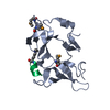
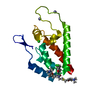
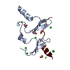
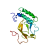
 Movie
Movie Controller


+ Open data

- Basic information
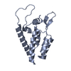
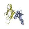
| Entry | Database: PDB / ID: 3tjq | ||||||
|---|---|---|---|---|---|---|---|
| Title | N-domain of HtrA1 | ||||||
 Components Components | Serine protease HTRA1 | ||||||
 Keywords Keywords |  HYDROLASE HYDROLASE | ||||||
| Function / homology |  Function and homology information Function and homology informationchorionic trophoblast cell differentiation / programmed cell death / growth factor binding / negative regulation of BMP signaling pathway / Hydrolases; Acting on peptide bonds (peptidases); Serine endopeptidases / serine-type peptidase activity / Degradation of the extracellular matrix / molecular function activator activity / placenta development / negative regulation of transforming growth factor beta receptor signaling pathway ...chorionic trophoblast cell differentiation / programmed cell death / growth factor binding / negative regulation of BMP signaling pathway / Hydrolases; Acting on peptide bonds (peptidases); Serine endopeptidases / serine-type peptidase activity / Degradation of the extracellular matrix / molecular function activator activity / placenta development / negative regulation of transforming growth factor beta receptor signaling pathway / collagen-containing extracellular matrix / positive regulation of apoptotic process / serine-type endopeptidase activity / proteolysis / extracellular space / extracellular exosome / extracellular region / identical protein binding / plasma membrane / cytosolSimilarity search - Function | ||||||
| Biological species |  Homo sapiens (human) Homo sapiens (human) | ||||||
| Method | X-RAY DIFFRACTION / SYNCHROTRON / SAD / Resolution: 2.001 Å | ||||||
 Authors Authors | Eigenbrot, C. / Ultsch, M. | ||||||
 Citation Citation | Journal: Structure / Year: 2012 Title: Structural and Functional Analysis of HtrA1 and Its Subdomains. Authors: Eigenbrot, C. / Ultsch, M. / Lipari, M.T. / Moran, P. / Lin, S.J. / Ganesan, R. / Quan, C. / Tom, J. / Sandoval, W. / van Lookeren Campagne, M. / Kirchhofer, D. | ||||||
| History |
|
- Structure visualization
| Structure viewer | Molecule: MolmilJmol/JSmol |
|---|
- Downloads & links
-Download
| PDBx/mmCIF format | 3tjq.cif.gz | 50.5 KB | Display | PDBx/mmCIF format |
|---|---|---|---|---|
| PDB format | pdb3tjq.ent.gz | 39.9 KB | Display | PDB format |
| PDBx/mmJSON format | 3tjq.json.gz | Tree view | PDBx/mmJSON format | |
| Others |  Other downloads Other downloads |
-Validation report
| Arichive directory | https://data.pdbj.org/pub/pdb/validation_reports/tj/3tjqftp://data.pdbj.org/pub/pdb/validation_reports/tj/3tjq | HTTPS FTP |
|---|
-Related structure data


-Links
 PDBj
PDBj


- Assembly
| Deposited unit | 
| ||||||||
|---|---|---|---|---|---|---|---|---|---|
| 1 |
| ||||||||
| Unit cell |
|
-Components
-Protein , 1 types, 1 molecules A
| #1: Protein | Mass: 14651.489 Da / Num. of mol.: 1 / Fragment: N-terminal part (UNP residues 36-155) Source method: isolated from a genetically manipulated source Source: (gene. exp.) Homo sapiens (human) / Gene: HTRA, HTRA1, PRSS11 / Production host:   Spodoptera frugiperda (fall armyworm) / Strain (production host): T. ni Spodoptera frugiperda (fall armyworm) / Strain (production host): T. niReferences: UniProt: Q92743, Hydrolases; Acting on peptide bonds (peptidases); Serine endopeptidases |
|---|
-Non-polymers , 5 types, 51 molecules 


| #2: Chemical |  Mass: 195.078 Da / Num. of mol.: 2 / Source method: obtained synthetically / Formula: Pt Mass: 195.078 Da / Num. of mol.: 2 / Source method: obtained synthetically / Formula: Pt#3: Chemical | ChemComp-CL / Chloride Mass: 35.453 Da / Num. of mol.: 6 / Source method: obtained synthetically / Formula: Cl Mass: 35.453 Da / Num. of mol.: 6 / Source method: obtained synthetically / Formula: Cl#4: Chemical | ChemComp-SCN / Thiocyanate Mass: 58.082 Da / Num. of mol.: 4 / Source method: obtained synthetically / Formula: CNS Mass: 58.082 Da / Num. of mol.: 4 / Source method: obtained synthetically / Formula: CNS#5: Chemical | ChemComp-GOL / | Glycerol Mass: 92.094 Da / Num. of mol.: 1 / Source method: obtained synthetically / Formula: C3H8O3 Mass: 92.094 Da / Num. of mol.: 1 / Source method: obtained synthetically / Formula: C3H8O3#6: Water | ChemComp-HOH / | WaterMass: 18.015 Da / Num. of mol.: 38 / Source method: isolated from a natural source / Formula: H2O |
|---|
-Experimental details
-Experiment
| Experiment | Method: X-RAY DIFFRACTION / Number of used crystals: 1 |
|---|
- Sample preparation
| Crystal | Density Matthews: 2.11 Å3/Da / Density % sol: 41.81 % |
|---|---|
| Crystal grow | Temperature: 300 K / Method: vapor diffusion, sitting drop / pH: 4.5 Details: potassium thiocyanate, sodium acetate, pH 4.5, VAPOR DIFFUSION, SITTING DROP, temperature 300K |
-Data collection
| Diffraction | Mean temperature: 110 K |
|---|---|
| Diffraction source | Source: SYNCHROTRON / Site: ALS  / Beamline: 5.0.1 / Wavelength: 0.98 Å / Beamline: 5.0.1 / Wavelength: 0.98 Å |
| Detector | Type: ADSC QUANTUM 315r / Detector: CCD / Date: Nov 23, 2010 |
| Radiation | Protocol: SINGLE WAVELENGTH / Monochromatic (M) / Laue (L): M / Scattering type: x-ray |
| Radiation wavelength | Wavelength: 0.98 Å / Relative weight: 1 |
| Reflection | Resolution: 2→50 Å / Num. all: 8555 / Num. obs: 7676 / % possible obs: 90 % / Observed criterion σ(F): 0 / Observed criterion σ(I): -2 / Redundancy: 9.8 % / Rsym value: 0.106 / Net I/σ(I): 19 |
- Processing
| Software |
| |||||||||||||||||||||||||||||||||||||||||||||||||||||||||||||||||||||||||||
|---|---|---|---|---|---|---|---|---|---|---|---|---|---|---|---|---|---|---|---|---|---|---|---|---|---|---|---|---|---|---|---|---|---|---|---|---|---|---|---|---|---|---|---|---|---|---|---|---|---|---|---|---|---|---|---|---|---|---|---|---|---|---|---|---|---|---|---|---|---|---|---|---|---|---|---|---|
| Refinement | Method to determine structure: SAD / Resolution: 2.001→40 Å / Cor.coef. Fo:Fc: 0.941 / Cor.coef. Fo:Fc free: 0.91 / SU B: 7.584 / SU ML: 0.119 / Cross valid method: THROUGHOUT / σ(F): 0 / σ(I): 0 / ESU R Free: 0.184 / Stereochemistry target values: MAXIMUM LIKELIHOOD / Details: HYDROGENS HAVE BEEN ADDED IN THE RIDING POSITIONS
| |||||||||||||||||||||||||||||||||||||||||||||||||||||||||||||||||||||||||||
| Solvent computation | Ion probe radii: 0.8 Å / Shrinkage radii: 0.8 Å / VDW probe radii: 1.4 Å / Solvent model: MASK | |||||||||||||||||||||||||||||||||||||||||||||||||||||||||||||||||||||||||||
| Displacement parameters | Biso mean: 41.153 Å2
| |||||||||||||||||||||||||||||||||||||||||||||||||||||||||||||||||||||||||||
| Refinement step | Cycle: LAST / Resolution: 2.001→40 Å
| |||||||||||||||||||||||||||||||||||||||||||||||||||||||||||||||||||||||||||
| Refine LS restraints |
| |||||||||||||||||||||||||||||||||||||||||||||||||||||||||||||||||||||||||||
| LS refinement shell | Resolution: 2.001→2.109 Å / Total num. of bins used: 10
| |||||||||||||||||||||||||||||||||||||||||||||||||||||||||||||||||||||||||||
| Refinement TLS params. | Method: refined / Refine-ID: X-RAY DIFFRACTION
| |||||||||||||||||||||||||||||||||||||||||||||||||||||||||||||||||||||||||||
| Refinement TLS group |
|
