 Movie
Movie Controller

[English] 日本語
 Yorodumi
- PDB-3th6: Crystal structure of Triosephosphate isomerase from Rhipicephalus... -
+ Open data

- Basic information
| Entry | Database: PDB / ID: 3th6 | ||||||
|---|---|---|---|---|---|---|---|
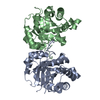
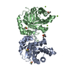
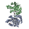
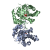
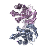
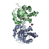
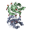
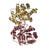
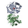
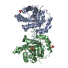
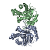
| Title | Crystal structure of Triosephosphate isomerase from Rhipicephalus (Boophilus) microplus. | ||||||
 Components Components | Triosephosphate isomerase | ||||||
 Keywords Keywords | ISOMERASE / ALPHA/BETA BARREL / EMBRYOGENESIS / GLYCOLYSIS | ||||||
| Function / homology |  Function and homology informationtriose-phosphate isomerase / triose-phosphate isomerase activity / gluconeogenesis / glycolytic process Function and homology informationtriose-phosphate isomerase / triose-phosphate isomerase activity / gluconeogenesis / glycolytic processSimilarity search - Function | ||||||
| Biological species |  Rhipicephalus microplus (southern cattle tick) Rhipicephalus microplus (southern cattle tick) | ||||||
| Method | X-RAY DIFFRACTION / SYNCHROTRON / MOLECULAR REPLACEMENT / molecular replacement / Resolution: 2.4 Å | ||||||
 Authors Authors | Arreola, R. / Rodriguez-Romero, A. / Moraes, J. / Gomez-Puyou, A. / Perez-Montfort, R. / Logullo, C. | ||||||
 Citation Citation | Journal: Insect Biochem.Mol.Biol. / Year: 2011 Title: Structural and biochemical characterization of a recombinant triosephosphate isomerase from Rhipicephalus (Boophilus) microplus. Authors: Moraes, J. / Arreola, R. / Cabrera, N. / Saramago, L. / Freitas, D. / Masuda, A. / da Silva Vaz, I. / Tuena de Gomez-Puyou, M. / Perez-Montfort, R. / Gomez-Puyou, A. / Logullo, C. | ||||||
| History |
|
- Structure visualization
| Structure viewer | Molecule: MolmilJmol/JSmol |
|---|
- Downloads & links
-Download
| PDBx/mmCIF format | 3th6.cif.gz | 105.5 KB | Display | PDBx/mmCIF format |
|---|---|---|---|---|
| PDB format | pdb3th6.ent.gz | 80.6 KB | Display | PDB format |
| PDBx/mmJSON format | 3th6.json.gz | Tree view | PDBx/mmJSON format | |
| Others |  Other downloads Other downloads |
-Validation report
| Arichive directory | https://data.pdbj.org/pub/pdb/validation_reports/th/3th6ftp://data.pdbj.org/pub/pdb/validation_reports/th/3th6 | HTTPS FTP |
|---|
-Related structure data
| Related structure data | 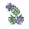 1wyiS S: Starting model for refinement |
|---|---|
| Similar structure data |


-Links
 PDBj
PDBj
- Assembly
| Deposited unit | 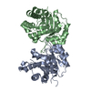
| ||||||||
|---|---|---|---|---|---|---|---|---|---|
| 1 |
| ||||||||
| Unit cell |
|
-Components
| #1: Protein | Mass: 27114.059 Da / Num. of mol.: 2 Source method: isolated from a genetically manipulated source Details: Total RNA was isolated from 10-day-old eggs Source: (gene. exp.) Rhipicephalus microplus (southern cattle tick)Strain: Porto Alegre / Gene: TIM / Plasmid: PET3A / Production host:  Escherichia coli (E. coli) / Strain (production host): BL21(DE3)PLYS / References: UniProt: A8B3A8, triose-phosphate isomerase Escherichia coli (E. coli) / Strain (production host): BL21(DE3)PLYS / References: UniProt: A8B3A8, triose-phosphate isomerase#2: Water | ChemComp-HOH / | Water Mass: 18.015 Da / Num. of mol.: 112 / Source method: isolated from a natural source / Formula: H2O Mass: 18.015 Da / Num. of mol.: 112 / Source method: isolated from a natural source / Formula: H2O |
|---|
-Experimental details
-Experiment
| Experiment | Method: X-RAY DIFFRACTION / Number of used crystals: 1 |
|---|
- Sample preparation
| Crystal | Density Matthews: 2.11 Å3/Da / Density % sol: 41.83 % |
|---|---|
| Crystal grow | Temperature: 291 K / Method: vapor diffusion, hanging drop / pH: 7.5 Details: 0.1 M HEPES/Na, 1.4 M Trisodium citrate dihydrate, pH 7.5, VAPOR DIFFUSION, HANGING DROP, temperature 291K |
-Data collection
| Diffraction | Mean temperature: 100 K | ||||||||||||||||||||||||||||||||||||||||||||||||||||||||||||||||||||||
|---|---|---|---|---|---|---|---|---|---|---|---|---|---|---|---|---|---|---|---|---|---|---|---|---|---|---|---|---|---|---|---|---|---|---|---|---|---|---|---|---|---|---|---|---|---|---|---|---|---|---|---|---|---|---|---|---|---|---|---|---|---|---|---|---|---|---|---|---|---|---|---|
| Diffraction source | Source: SYNCHROTRON / Site: APS  / Beamline: 5ID-B / Wavelength: 0.977 Å / Beamline: 5ID-B / Wavelength: 0.977 Å | ||||||||||||||||||||||||||||||||||||||||||||||||||||||||||||||||||||||
| Detector | Type: MAR CCD 165 mm / Detector: CCD / Date: Oct 5, 2006 | ||||||||||||||||||||||||||||||||||||||||||||||||||||||||||||||||||||||
| Radiation | Monochromator: Si(111) / Protocol: SINGLE WAVELENGTH / Monochromatic (M) / Laue (L): M / Scattering type: x-ray | ||||||||||||||||||||||||||||||||||||||||||||||||||||||||||||||||||||||
| Radiation wavelength | Wavelength: 0.977 Å / Relative weight: 1 | ||||||||||||||||||||||||||||||||||||||||||||||||||||||||||||||||||||||
| Reflection | Resolution: 2.4→46.474 Å / Num. all: 17583 / Num. obs: 17583 / % possible obs: 100 % / Observed criterion σ(F): 2 / Observed criterion σ(I): 2 / Redundancy: 8.5 % / Biso Wilson estimate: 35.9 Å2 / Rmerge(I) obs: 0.102 / Rsym value: 0.102 / Net I/σ(I): 17.3 | ||||||||||||||||||||||||||||||||||||||||||||||||||||||||||||||||||||||
| Reflection shell | Diffraction-ID: 1 / % possible all: 100
|
-Phasing
| Phasing | Method: molecular replacement | |||||||||
|---|---|---|---|---|---|---|---|---|---|---|
| Phasing MR | Model details: Phaser MODE: MR_AUTO
|
- Processing
| Software |
| ||||||||||||||||||||||||||||||||||||
|---|---|---|---|---|---|---|---|---|---|---|---|---|---|---|---|---|---|---|---|---|---|---|---|---|---|---|---|---|---|---|---|---|---|---|---|---|---|
| Refinement | Method to determine structure: MOLECULAR REPLACEMENT Starting model: PDB entry 1WYI Resolution: 2.4→46.46 Å / Rfactor Rfree error: 0.005 / Occupancy max: 1 / Occupancy min: 0 / Data cutoff high absF: 1907544 / Data cutoff low absF: 0 / Isotropic thermal model: RESTRAINED / Cross valid method: THROUGHOUT / σ(F): 0 / Stereochemistry target values: Engh & Huber Details: BULK SOLVENT MODEL USED THE FOLLOWING RESIDUES ARE DISORDERED AND IT WERE NOT MODELED DUE TO POOR/NULL ELECTRON DENSITY. CHAIN A/B RESIDUES: MET1 AND ALA2. CHAIN A RESIDUES FROM LOOP6: ...Details: BULK SOLVENT MODEL USED THE FOLLOWING RESIDUES ARE DISORDERED AND IT WERE NOT MODELED DUE TO POOR/NULL ELECTRON DENSITY. CHAIN A/B RESIDUES: MET1 AND ALA2. CHAIN A RESIDUES FROM LOOP6: THR172, GLY173 AND LYS174. CHAIN B RESIDUES FROM LOOP6: GLY171 THR172, GLY173, LYS174 AND THR175. CHAIN B RESIDUE: GLY249
| ||||||||||||||||||||||||||||||||||||
| Solvent computation | Solvent model: FLAT MODEL / Bsol: 42.5156 Å2 / ksol: 0.4 e/Å3 | ||||||||||||||||||||||||||||||||||||
| Displacement parameters | Biso max: 80.29 Å2 / Biso mean: 29.8919 Å2 / Biso min: 8.29 Å2
| ||||||||||||||||||||||||||||||||||||
| Refine analyze |
| ||||||||||||||||||||||||||||||||||||
| Refinement step | Cycle: LAST / Resolution: 2.4→46.46 Å
| ||||||||||||||||||||||||||||||||||||
| Refine LS restraints |
| ||||||||||||||||||||||||||||||||||||
| LS refinement shell | Resolution: 2.4→2.55 Å / Rfactor Rfree error: 0.018 / Total num. of bins used: 6
| ||||||||||||||||||||||||||||||||||||
| Xplor file |
|
