 Movie
Movie Controller

[English] 日本語
 Yorodumi
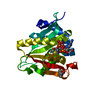
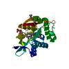
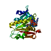
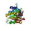
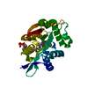
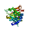
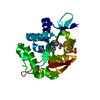
- PDB-3te4: Crystal structure of dopamine N Acetyltransferase in complex with... -
+ Open data

- Basic information
| Entry | Database: PDB / ID: 3te4 | ||||||
|---|---|---|---|---|---|---|---|
| Title | Crystal structure of dopamine N Acetyltransferase in complex with acetyl-COA from Drosophila Melanogaster | ||||||
 Components Components | Dopamine N acetyltransferase, isoform A | ||||||
 Keywords Keywords |  TRANSFERASE / DOPAMINE/ACETYL COA / N-acetyltransferase domain TRANSFERASE / DOPAMINE/ACETYL COA / N-acetyltransferase domain | ||||||
| Function / homology |  Function and homology information Function and homology informationchitin-based cuticle sclerotization / serotonin catabolic process / octopamine catabolic process / aralkylamine N-acetyltransferase / aralkylamine N-acetyltransferase activity / arylamine N-acetyltransferase activity / melatonin biosynthetic process / regulation of circadian sleep/wake cycle, sleep / catecholamine metabolic process / dopamine catabolic process ...chitin-based cuticle sclerotization / serotonin catabolic process / octopamine catabolic process / aralkylamine N-acetyltransferase / aralkylamine N-acetyltransferase activity / arylamine N-acetyltransferase activity / melatonin biosynthetic process / regulation of circadian sleep/wake cycle, sleep / catecholamine metabolic process / dopamine catabolic process / sleep / N-acetyltransferase activity / nucleus / cytoplasmSimilarity search - Function | ||||||
| Biological species |  Drosophila melanogaster (fruit fly) Drosophila melanogaster (fruit fly) | ||||||
| Method | X-RAY DIFFRACTION / SYNCHROTRON / SAD / Resolution: 1.46 Å | ||||||
 Authors Authors | Cheng, K.C. / Huang, S.H. / Lyu, P.C. | ||||||
 Citation Citation | Journal: Biochem.J. / Year: 2012 Title: Crystal structure of the dopamine N-acetyltransferase-acetyl-CoA complex provides insights into the catalytic mechanism Authors: Cheng, K.C. / Liao, J.N. / Lyu, P.C. | ||||||
| History |
|
- Structure visualization
| Structure viewer | Molecule: MolmilJmol/JSmol |
|---|
- Downloads & links
-Download
| PDBx/mmCIF format | 3te4.cif.gz | 114.6 KB | Display | PDBx/mmCIF format |
|---|---|---|---|---|
| PDB format | pdb3te4.ent.gz | 89.2 KB | Display | PDB format |
| PDBx/mmJSON format | 3te4.json.gz | Tree view | PDBx/mmJSON format | |
| Others |  Other downloads Other downloads |
-Validation report
| Arichive directory | https://data.pdbj.org/pub/pdb/validation_reports/te/3te4ftp://data.pdbj.org/pub/pdb/validation_reports/te/3te4 | HTTPS FTP |
|---|
-Related structure data
| Similar structure data |
|---|


-Links
 PDBj
PDBj

- Assembly
| Deposited unit | 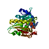
| ||||||||
|---|---|---|---|---|---|---|---|---|---|
| 1 |
| ||||||||
| Unit cell |
|
-Components
| #1: Protein | Mass: 24442.049 Da / Num. of mol.: 1 / Fragment: UNP residues 21-230 Source method: isolated from a genetically manipulated source Source: (gene. exp.) Drosophila melanogaster (fruit fly) / Gene: CG3318, Dat, Dmel_CG3318 / Plasmid: PGEX-6P-3 / Production host:  Escherichia coli (E. coli) / Strain (production host): BL21(DE3) Escherichia coli (E. coli) / Strain (production host): BL21(DE3)References: UniProt: Q8MKK2, UniProt: Q94521*PLUS, Transferases; Acyltransferases; Transferring groups other than aminoacyl groups, arylamine N-acetyltransferase, aralkylamine N-acetyltransferase |
|---|---|
| #2: Chemical | ChemComp-ACO / Acetyl-CoA  Mass: 809.571 Da / Num. of mol.: 1 / Source method: obtained synthetically / Formula: C23H38N7O17P3S Mass: 809.571 Da / Num. of mol.: 1 / Source method: obtained synthetically / Formula: C23H38N7O17P3S |
| #3: Water | ChemComp-HOH / Water Mass: 18.015 Da / Num. of mol.: 345 / Source method: isolated from a natural source / Formula: H2O Mass: 18.015 Da / Num. of mol.: 345 / Source method: isolated from a natural source / Formula: H2O |
-Experimental details
-Experiment
| Experiment | Method: X-RAY DIFFRACTION / Number of used crystals: 1 |
|---|
- Sample preparation
| Crystal | Density Matthews: 2.14 Å3/Da / Density % sol: 42.54 % |
|---|---|
| Crystal grow | Temperature: 277 K / Method: vapor diffusion, hanging drop / pH: 8.5 Details: 1.0M NAH2PO4/1.6M K2HPO4, 0.1M IMIDAZOLE, 0.2M NACL , pH 8.5, VAPOR DIFFUSION, HANGING DROP, temperature 277K |
-Data collection
| Diffraction | Mean temperature: 100 K |
|---|---|
| Diffraction source | Source: SYNCHROTRON / Site: NSRRC  / Beamline: BL13B1 / Wavelength: 0.96369 Å / Beamline: BL13B1 / Wavelength: 0.96369 Å |
| Detector | Type: ADSC QUANTUM 315r / Detector: CCD / Date: Jul 23, 2010 |
| Radiation | Protocol: SINGLE WAVELENGTH / Monochromatic (M) / Laue (L): M / Scattering type: x-ray |
| Radiation wavelength | Wavelength: 0.96369 Å / Relative weight: 1 |
| Reflection | Resolution: 1.46→30 Å / Num. obs: 36678 / % possible obs: 98.7 % / Redundancy: 5.5 % / Biso Wilson estimate: 19.1 Å2 / Rmerge(I) obs: 0.058 / Net I/σ(I): 23.4 |
| Reflection shell | Resolution: 1.46→1.51 Å / Redundancy: 5.3 % / Rmerge(I) obs: 0.205 / Mean I/σ(I) obs: 9.25 / % possible all: 98.7 |
- Processing
| Software |
| ||||||||||||||||||||||||||||||||||||||||||||||||||||||||||||||||||||||
|---|---|---|---|---|---|---|---|---|---|---|---|---|---|---|---|---|---|---|---|---|---|---|---|---|---|---|---|---|---|---|---|---|---|---|---|---|---|---|---|---|---|---|---|---|---|---|---|---|---|---|---|---|---|---|---|---|---|---|---|---|---|---|---|---|---|---|---|---|---|---|---|
| Refinement | Method to determine structure: SAD / Resolution: 1.46→23.61 Å / Cor.coef. Fo:Fc: 0.969 / Cor.coef. Fo:Fc free: 0.948 / SU B: 2.139 / SU ML: 0.038 / Cross valid method: THROUGHOUT / ESU R Free: 0.072 / Stereochemistry target values: MAXIMUM LIKELIHOOD Details: HYDROGENS HAVE BEEN ADDED IN THE RIDING POSITIONS U VALUES: REFINED INDIVIDUALLY
| ||||||||||||||||||||||||||||||||||||||||||||||||||||||||||||||||||||||
| Solvent computation | Ion probe radii: 0.8 Å / Shrinkage radii: 0.8 Å / VDW probe radii: 1.4 Å / Solvent model: MASK | ||||||||||||||||||||||||||||||||||||||||||||||||||||||||||||||||||||||
| Displacement parameters | Biso mean: 16.4 Å2
| ||||||||||||||||||||||||||||||||||||||||||||||||||||||||||||||||||||||
| Refinement step | Cycle: LAST / Resolution: 1.46→23.61 Å
| ||||||||||||||||||||||||||||||||||||||||||||||||||||||||||||||||||||||
| Refine LS restraints |
| ||||||||||||||||||||||||||||||||||||||||||||||||||||||||||||||||||||||
| LS refinement shell | Resolution: 1.46→1.5 Å / Total num. of bins used: 20
|
