- PDB-3tb3: Crystal structure of the UCH domain of UCH-L5 with 6 residues deleted -
+
Open data
ID or keywords:
Loading...
-
Basic information
Entry
Database: PDB / ID: 3tb3
Title
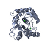
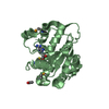
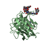
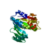
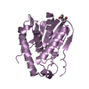
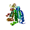
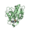
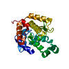
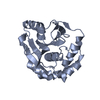
Crystal structure of the UCH domain of UCH-L5 with 6 residues deleted
Components
Ubiquitin carboxyl-terminal hydrolase isozyme L5
Keywords
HYDROLASE / UCH domain
Function / homology
Function and homology information
lateral ventricle development / regulation of DNA strand elongation / positive regulation of telomere maintenance in response to DNA damage / forebrain morphogenesis / Ino80 complex / cytosolic proteasome complex / positive regulation of smoothened signaling pathway / midbrain development / regulation of chromosome organization / endopeptidase inhibitor activity ...lateral ventricle development / regulation of DNA strand elongation / positive regulation of telomere maintenance in response to DNA damage / forebrain morphogenesis / Ino80 complex / cytosolic proteasome complex / positive regulation of smoothened signaling pathway / midbrain development / regulation of chromosome organization / endopeptidase inhibitor activity / proteasome binding / protein deubiquitination / regulation of DNA replication / regulation of embryonic development / negative regulation of proteasomal ubiquitin-dependent protein catabolic process / regulation of DNA repair / regulation of proteasomal protein catabolic process / positive regulation of DNA repair / telomere maintenance / Downregulation of TGF-beta receptor signaling / UCH proteinases / ubiquitin-dependent protein catabolic process / DNA recombination / ubiquitinyl hydrolase 1 / cysteine-type deubiquitinase activity / regulation of cell cycle / chromatin remodeling / DNA repair / nucleolus / positive regulation of DNA-templated transcription / RNA binding / nucleoplasm / nucleus / cytosol / cytoplasm Similarity search - Function
In the structure databanks used in Yorodumi, some data are registered as the other names, "COVID-19 virus" and "2019-nCoV". Here are the details of the virus and the list of structure data.
Jan 31, 2019. EMDB accession codes are about to change! (news from PDBe EMDB page)
EMDB accession codes are about to change! (news from PDBe EMDB page)
The allocation of 4 digits for EMDB accession codes will soon come to an end. Whilst these codes will remain in use, new EMDB accession codes will include an additional digit and will expand incrementally as the available range of codes is exhausted. The current 4-digit format prefixed with “EMD-” (i.e. EMD-XXXX) will advance to a 5-digit format (i.e. EMD-XXXXX), and so on. It is currently estimated that the 4-digit codes will be depleted around Spring 2019, at which point the 5-digit format will come into force.
The EM Navigator/Yorodumi systems omit the EMD- prefix.
Related info.:Q: What is EMD? / ID/Accession-code notation in Yorodumi/EM Navigator
Yorodumi is a browser for structure data from EMDB, PDB, SASBDB, etc.
This page is also the successor to EM Navigator detail page, and also detail information page/front-end page for Omokage search.
The word "yorodu" (or yorozu) is an old Japanese word meaning "ten thousand". "mi" (miru) is to see.
Related info.:EMDB / PDB / SASBDB / Comparison of 3 databanks / Yorodumi Search / Aug 31, 2016. New EM Navigator & Yorodumi / Yorodumi Papers / Jmol/JSmol / Function and homology information / Changes in new EM Navigator and Yorodumi
 Movie
Movie Controller

 Yorodumi
 Open data
 Basic information
 Components
 Keywords
 HYDROLASE / UCH domain
 Function and homology information
 Authors
 Citation
 Structure visualization
 Downloads & links
 Other downloads
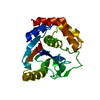


 PDBj
PDBj


 Assembly


 Mass: 40.078 Da / Num. of mol.: 2 / Source method: obtained synthetically / Formula: Ca
 Mass: 18.015 Da / Num. of mol.: 43 / Source method: isolated from a natural source / Formula: H2O
 Sample preparation
 / Beamline: BL17U / Wavelength: 0.9791 Å
 Processing