 Movie
Movie Controller

[English] 日本語
 Yorodumi
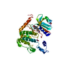
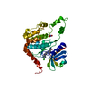
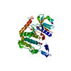
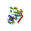
- PDB-3t3a: Crystal structure of H107R mutant of extracellular domain of mous... -
+ Open data

- Basic information
| Entry | Database: PDB / ID: 3t3a | ||||||
|---|---|---|---|---|---|---|---|
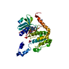
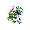
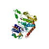
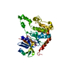
| Title | Crystal structure of H107R mutant of extracellular domain of mouse receptor NKR-P1A | ||||||
 Components Components | Killer cell lectin-like receptor subfamily B member 1A | ||||||
 Keywords Keywords |  SIGNALING PROTEIN / C-type lectin-like domain / domain swapping / twinning / natural killer cell receptor / transmembrane receptor SIGNALING PROTEIN / C-type lectin-like domain / domain swapping / twinning / natural killer cell receptor / transmembrane receptor | ||||||
| Function / homology |  Function and homology information Function and homology informationregulation of natural killer cell mediated cytotoxicity / signaling receptor activity / carbohydrate binding / cell surface / plasma membraneSimilarity search - Function | ||||||
| Biological species |  Mus musculus (house mouse) Mus musculus (house mouse) | ||||||
| Method | X-RAY DIFFRACTION / SYNCHROTRON / MOLECULAR REPLACEMENT / Resolution: 2.3 Å | ||||||
 Authors Authors | Kolenko, P. / Rozbesky, D. / Bezouska, K. / Hasek, J. / Dohnalek, J. | ||||||
 Citation Citation | Journal: Acta Crystallogr.,Sect.F / Year: 2011 Title: Structure of the H107R variant of the extracellular domain of mouse NKR-P1A at 2.3 A resolution. Authors: Kolenko, P. / Rozbesky, D. / Vanek, O. / Bezouska, K. / Hasek, J. / Dohnalek, J. | ||||||
| History |
|
- Structure visualization
| Structure viewer | Molecule: MolmilJmol/JSmol |
|---|
- Downloads & links
-Download
| PDBx/mmCIF format | 3t3a.cif.gz | 63.3 KB | Display | PDBx/mmCIF format |
|---|---|---|---|---|
| PDB format | pdb3t3a.ent.gz | 46.6 KB | Display | PDB format |
| PDBx/mmJSON format | 3t3a.json.gz | Tree view | PDBx/mmJSON format | |
| Others |  Other downloads Other downloads |
-Validation report
| Arichive directory | https://data.pdbj.org/pub/pdb/validation_reports/t3/3t3aftp://data.pdbj.org/pub/pdb/validation_reports/t3/3t3a | HTTPS FTP |
|---|
-Related structure data
| Related structure data |  3m9zS S: Starting model for refinement |
|---|---|
| Similar structure data |
-Links
 PDBj
PDBj


- Assembly
| Deposited unit | 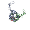
| ||||||||
|---|---|---|---|---|---|---|---|---|---|
| 1 |
| ||||||||
| 2 | 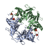
| ||||||||
| Unit cell |
| ||||||||
| Components on special symmetry positions |
|
-Components
| #1: Protein | Mass: 16027.820 Da / Num. of mol.: 2 / Fragment: Extracellular domain (UNP residues 89-227) / Mutation: H107R Source method: isolated from a genetically manipulated source Source: (gene. exp.) Mus musculus (house mouse) / Strain: C57BL/6 / Gene: Klrb1a, Ly55, Ly55a, Nkrp1a / Plasmid: PET-30A / Production host:  Escherichia coli (E. coli) / Strain (production host): BL21-(DE3) RIL / References: UniProt: P27811 Escherichia coli (E. coli) / Strain (production host): BL21-(DE3) RIL / References: UniProt: P27811#2: Chemical | Phosphate  Mass: 94.971 Da / Num. of mol.: 3 / Source method: obtained synthetically / Formula: PO4 Mass: 94.971 Da / Num. of mol.: 3 / Source method: obtained synthetically / Formula: PO4#3: Water | ChemComp-HOH / | Water Mass: 18.015 Da / Num. of mol.: 56 / Source method: isolated from a natural source / Formula: H2O Mass: 18.015 Da / Num. of mol.: 56 / Source method: isolated from a natural source / Formula: H2O |
|---|
-Experimental details
-Experiment
| Experiment | Method: X-RAY DIFFRACTION / Number of used crystals: 1 |
|---|
- Sample preparation
| Crystal | Density Matthews: 2.55 Å3/Da / Density % sol: 51.79 % |
|---|---|
| Crystal grow | Temperature: 291 K / Method: vapor diffusion, hanging drop / pH: 4.2 Details: 0.3M ammonium phosphate, pH 4.2, VAPOR DIFFUSION, HANGING DROP, temperature 291K |
-Data collection
| Diffraction | Mean temperature: 100 K | |||||||||||||||||||||
|---|---|---|---|---|---|---|---|---|---|---|---|---|---|---|---|---|---|---|---|---|---|---|
| Diffraction source | Source: SYNCHROTRON / Site: BESSY  / Beamline: 14.1 / Wavelength: 0.91841 Å / Beamline: 14.1 / Wavelength: 0.91841 Å | |||||||||||||||||||||
| Detector | Type: MARMOSAIC 225 mm CCD / Detector: CCD / Date: May 12, 2009 / Details: 2 mirrors and a double-crystal monochromator | |||||||||||||||||||||
| Radiation | Monochromator: KMC-1, Si (111) / Protocol: SINGLE WAVELENGTH / Monochromatic (M) / Laue (L): M / Scattering type: x-ray | |||||||||||||||||||||
| Radiation wavelength | Wavelength: 0.91841 Å / Relative weight: 1 | |||||||||||||||||||||
| Reflection twin |
| |||||||||||||||||||||
| Reflection | Resolution: 2.3→60 Å / Num. all: 14227 / Num. obs: 14227 / % possible obs: 99.9 % / Observed criterion σ(F): 0 / Observed criterion σ(I): -3.7 / Redundancy: 7.5 % / Biso Wilson estimate: 49 Å2 / Rmerge(I) obs: 0.072 / Net I/σ(I): 16 | |||||||||||||||||||||
| Reflection shell |
|
- Processing
| Software |
| ||||||||||||||||||||||||||||||||||||||||||||||||||||||||||||||||||||||||||||||||||||||||||||||||||||||||||||||||||||||||||||||||||||||||||||||||||||||||||||||||||||||||||
|---|---|---|---|---|---|---|---|---|---|---|---|---|---|---|---|---|---|---|---|---|---|---|---|---|---|---|---|---|---|---|---|---|---|---|---|---|---|---|---|---|---|---|---|---|---|---|---|---|---|---|---|---|---|---|---|---|---|---|---|---|---|---|---|---|---|---|---|---|---|---|---|---|---|---|---|---|---|---|---|---|---|---|---|---|---|---|---|---|---|---|---|---|---|---|---|---|---|---|---|---|---|---|---|---|---|---|---|---|---|---|---|---|---|---|---|---|---|---|---|---|---|---|---|---|---|---|---|---|---|---|---|---|---|---|---|---|---|---|---|---|---|---|---|---|---|---|---|---|---|---|---|---|---|---|---|---|---|---|---|---|---|---|---|---|---|---|---|---|---|---|---|
| Refinement | Method to determine structure: MOLECULAR REPLACEMENT Starting model: 3M9Z Resolution: 2.3→50 Å / Cor.coef. Fo:Fc: 0.952 / Cor.coef. Fo:Fc free: 0 / SU B: 2.411 / SU ML: 0.062 / Isotropic thermal model: Isotropic / Cross valid method: THROUGHOUT / σ(F): 0 Stereochemistry target values: CCP4 stereochemistry library, version 6.1.3 Details: INTENSITY-BASED TWIN REFINEMENT, ALPHA(TWIN) 14%, HYDROGENS HAVE BEEN ADDED IN THE RIDING POSITIONS
| ||||||||||||||||||||||||||||||||||||||||||||||||||||||||||||||||||||||||||||||||||||||||||||||||||||||||||||||||||||||||||||||||||||||||||||||||||||||||||||||||||||||||||
| Solvent computation | Ion probe radii: 0.8 Å / Shrinkage radii: 0.8 Å / VDW probe radii: 1.2 Å / Solvent model: MASK | ||||||||||||||||||||||||||||||||||||||||||||||||||||||||||||||||||||||||||||||||||||||||||||||||||||||||||||||||||||||||||||||||||||||||||||||||||||||||||||||||||||||||||
| Displacement parameters | Biso mean: 37.893 Å2
| ||||||||||||||||||||||||||||||||||||||||||||||||||||||||||||||||||||||||||||||||||||||||||||||||||||||||||||||||||||||||||||||||||||||||||||||||||||||||||||||||||||||||||
| Refinement step | Cycle: LAST / Resolution: 2.3→50 Å
| ||||||||||||||||||||||||||||||||||||||||||||||||||||||||||||||||||||||||||||||||||||||||||||||||||||||||||||||||||||||||||||||||||||||||||||||||||||||||||||||||||||||||||
| Refine LS restraints |
| ||||||||||||||||||||||||||||||||||||||||||||||||||||||||||||||||||||||||||||||||||||||||||||||||||||||||||||||||||||||||||||||||||||||||||||||||||||||||||||||||||||||||||
| LS refinement shell | Resolution: 2.299→2.359 Å / Total num. of bins used: 20
|
