 Movie
Movie Controller

[English] 日本語
 Yorodumi
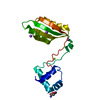
- PDB-3sxl: SEX-LETHAL RNA RECOGNITION DOMAINS 1 AND 2 FROM DROSOPHILA MELANO... -
+ Open data

- Basic information
| Entry | Database: PDB / ID: 3sxl | ||||||
|---|---|---|---|---|---|---|---|
| Title | SEX-LETHAL RNA RECOGNITION DOMAINS 1 AND 2 FROM DROSOPHILA MELANOGASTER | ||||||
 Components Components | PROTEIN (SEX-LETHAL) | ||||||
 Keywords Keywords | RNA BINDING DOMAIN / RBD /  RNA RECOGNITION MOTIF / RRM / SPLICING INHIBITOR / TRANSLATIONAL INHIBITOR / SEX DETERMINATION / X CHROMOSOME DOSAGE COMPENSATION RNA RECOGNITION MOTIF / RRM / SPLICING INHIBITOR / TRANSLATIONAL INHIBITOR / SEX DETERMINATION / X CHROMOSOME DOSAGE COMPENSATION | ||||||
| Function / homology |  Function and homology information Function and homology informationsex determination, primary response to X:A ratio / germarium-derived cystoblast division / epithelium regeneration / female sex determination / somatic sex determination / female germ-line sex determination / oocyte differentiation / imaginal disc growth / negative regulation of RNA export from nucleus / sex-chromosome dosage compensation ...sex determination, primary response to X:A ratio / germarium-derived cystoblast division / epithelium regeneration / female sex determination / somatic sex determination / female germ-line sex determination / oocyte differentiation / imaginal disc growth / negative regulation of RNA export from nucleus / sex-chromosome dosage compensation / regulation of stem cell division / sex determination / poly-pyrimidine tract binding / sex differentiation / alternative mRNA splicing, via spliceosome / negative regulation of receptor signaling pathway via JAK-STAT / poly(A) binding / pre-mRNA binding / positive regulation of smoothened signaling pathway / regulation of mRNA splicing, via spliceosome / reciprocal meiotic recombination / poly(U) RNA binding / oogenesis / regulation of alternative mRNA splicing, via spliceosome / negative regulation of mRNA splicing, via spliceosome / negative regulation of translational initiation / mRNA regulatory element binding translation repressor activity / positive regulation of RNA splicing / mRNA 3'-UTR binding / mRNA 5'-UTR binding / negative regulation of translation / protein stabilization / ribonucleoprotein complex / mRNA binding / protein-containing complex / RNA binding / nucleus / cytosol / cytoplasmSimilarity search - Function | ||||||
| Biological species |  Drosophila melanogaster (fruit fly) Drosophila melanogaster (fruit fly) | ||||||
| Method | X-RAY DIFFRACTION / SYNCHROTRON / MAD / Resolution: 2.7 Å | ||||||
 Authors Authors | Crowder, S.M. / Kanaar, R. / Rio, D.C. / Alber, T.C. | ||||||
 Citation Citation | Journal: Proc.Natl.Acad.Sci.USA / Year: 1999 Title: Absence of interdomain contacts in the crystal structure of the RNA recognition motifs of Sex-lethal. Authors: Crowder, S.M. / Kanaar, R. / Rio, D.C. / Alber, T. | ||||||
| History |
|
- Structure visualization
| Structure viewer | Molecule: MolmilJmol/JSmol |
|---|
- Downloads & links
-Download
| PDBx/mmCIF format | 3sxl.cif.gz | 102.7 KB | Display | PDBx/mmCIF format |
|---|---|---|---|---|
| PDB format | pdb3sxl.ent.gz | 79.9 KB | Display | PDB format |
| PDBx/mmJSON format | 3sxl.json.gz | Tree view | PDBx/mmJSON format | |
| Others |  Other downloads Other downloads |
-Validation report
| Arichive directory | https://data.pdbj.org/pub/pdb/validation_reports/sx/3sxlftp://data.pdbj.org/pub/pdb/validation_reports/sx/3sxl | HTTPS FTP |
|---|
-Related structure data
| Similar structure data |
|---|


-Links
 PDBj
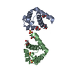
PDBj- Assembly
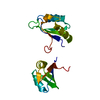
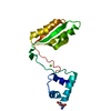
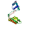
| Deposited unit | 
| ||||||||
|---|---|---|---|---|---|---|---|---|---|
| 1 | 
| ||||||||
| 2 | 
| ||||||||
| 3 | 
| ||||||||
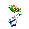
| Unit cell |
| ||||||||
| Noncrystallographic symmetry (NCS) | NCS oper: (Code: given / Matrix: (1, 1),: |
-Components
| #1: Protein | Mass: 21031.182 Da / Num. of mol.: 3 Fragment: RNA BINDING DOMAINS 1 AND 2 (RESIDUES 112-294 OF FULL LENGTH) Source method: isolated from a genetically manipulated source Source: (gene. exp.) Drosophila melanogaster (fruit fly) / Plasmid: PET-3B / Species (production host): Escherichia coli / Production host:  Escherichia coli BL21(DE3) (bacteria) / Strain (production host): BL-21(DE3) / References: UniProt: P19339 Escherichia coli BL21(DE3) (bacteria) / Strain (production host): BL-21(DE3) / References: UniProt: P19339 |
|---|
-Experimental details
-Experiment
| Experiment | Method: X-RAY DIFFRACTION |
|---|
- Sample preparation
| Crystal | Density Matthews: 2.21 Å3/Da / Density % sol: 44.45 % | |||||||||||||||||||||||||||||||||||
|---|---|---|---|---|---|---|---|---|---|---|---|---|---|---|---|---|---|---|---|---|---|---|---|---|---|---|---|---|---|---|---|---|---|---|---|---|
| Crystal grow | pH: 7.8 / Details: pH 7.80 | |||||||||||||||||||||||||||||||||||
| Crystal grow | *PLUS pH: 7.8 / Method: vapor diffusion, hanging drop / Details: used to seeding | |||||||||||||||||||||||||||||||||||
| Components of the solutions | *PLUS
|
-Data collection
| Diffraction | Mean temperature: 120 K | |||||||||||||||
|---|---|---|---|---|---|---|---|---|---|---|---|---|---|---|---|---|
| Diffraction source | Source: SYNCHROTRON / Site: SSRL  / Beamline: BL1-5 / Wavelength: 1.06879, 0.98001, 0.97957, 1.2000 / Beamline: BL1-5 / Wavelength: 1.06879, 0.98001, 0.97957, 1.2000 | |||||||||||||||
| Detector | Detector: CCD | |||||||||||||||
| Radiation | Protocol: MAD / Monochromatic (M) / Laue (L): M / Scattering type: x-ray | |||||||||||||||
| Radiation wavelength |
| |||||||||||||||
| Reflection | Resolution: 2.7→20 Å / Num. obs: 16847 / % possible obs: 99.8 % / Observed criterion σ(I): 2 / Redundancy: 6.7 % / Biso Wilson estimate: 68.5 Å2 / Rsym value: 0.069 / Net I/σ(I): 13.4 | |||||||||||||||
| Reflection shell | Highest resolution: 2.7 Å / Mean I/σ(I) obs: 5.7 | |||||||||||||||
| Reflection | *PLUS Highest resolution: 2.7 Å / Lowest resolution: 20 Å / % possible obs: 99.8 % / Observed criterion σ(I): 2 / Redundancy: 6.7 % / Rmerge(I) obs: 0.069 / Biso Wilson estimate: 68.5 Å2 | |||||||||||||||
| Reflection shell | *PLUS Highest resolution: 2.7 Å / Mean I/σ(I) obs: 5.7 |
- Processing
| Software |
| ||||||||||||||||||||||||||||||||||||||||||||||||||||||||||||
|---|---|---|---|---|---|---|---|---|---|---|---|---|---|---|---|---|---|---|---|---|---|---|---|---|---|---|---|---|---|---|---|---|---|---|---|---|---|---|---|---|---|---|---|---|---|---|---|---|---|---|---|---|---|---|---|---|---|---|---|---|---|
| Refinement | Method to determine structure: MAD / Resolution: 2.7→20 Å / Rfactor Rfree error: 0.007 / Data cutoff high rms absF: 1604575.59 / Isotropic thermal model: RESTRAINED / Cross valid method: THROUGHOUT / σ(F): 2
| ||||||||||||||||||||||||||||||||||||||||||||||||||||||||||||
| Solvent computation | Solvent model: FLAT MODEL | ||||||||||||||||||||||||||||||||||||||||||||||||||||||||||||
| Displacement parameters | Biso mean: 56.5 Å2
| ||||||||||||||||||||||||||||||||||||||||||||||||||||||||||||
| Refine analyze |
| ||||||||||||||||||||||||||||||||||||||||||||||||||||||||||||
| Refinement step | Cycle: LAST / Resolution: 2.7→20 Å
| ||||||||||||||||||||||||||||||||||||||||||||||||||||||||||||
| Refine LS restraints |
| ||||||||||||||||||||||||||||||||||||||||||||||||||||||||||||
| LS refinement shell | Resolution: 2.7→2.87 Å / Rfactor Rfree error: 0.03 / Total num. of bins used: 6
| ||||||||||||||||||||||||||||||||||||||||||||||||||||||||||||
| Xplor file | Serial no: 1 / Param file: PROTEIN_REP.PARAM / Topol file: PROTEIN.TOP | ||||||||||||||||||||||||||||||||||||||||||||||||||||||||||||
| Refinement | *PLUS Highest resolution: 2.7 Å / Lowest resolution: 20 Å / σ(F): 2 / % reflection Rfree: 10 % / Rfactor obs: 0.218 | ||||||||||||||||||||||||||||||||||||||||||||||||||||||||||||
| Solvent computation | *PLUS | ||||||||||||||||||||||||||||||||||||||||||||||||||||||||||||
| Displacement parameters | *PLUS Biso mean: 56.5 Å2 | ||||||||||||||||||||||||||||||||||||||||||||||||||||||||||||
| Refine LS restraints | *PLUS
| ||||||||||||||||||||||||||||||||||||||||||||||||||||||||||||
| LS refinement shell | *PLUS Highest resolution: 2.7 Å / % reflection Rfree: 10.6 % |
