 Movie
Movie Controller


+ Open data

- Basic information
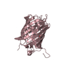
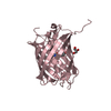
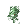
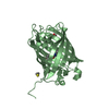
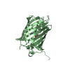
| Entry | Database: PDB / ID: 3svn | ||||||
|---|---|---|---|---|---|---|---|
| Title | Crystal structure of mKate S158A mutant at pH 7.5 | ||||||
 Components Components | mKate | ||||||
 Keywords Keywords |  FLUORESCENT PROTEIN / Fluoresent protein / pH sensor FLUORESCENT PROTEIN / Fluoresent protein / pH sensor | ||||||
| Function / homology | Green Fluorescent Protein / Green fluorescent protein / Beta Barrel / Mainly Beta Function and homology information Function and homology information | ||||||
| Biological species | Artificial gene (others) | ||||||
| Method | X-RAY DIFFRACTION / SYNCHROTRON / MOLECULAR REPLACEMENT / Resolution: 1.899 Å | ||||||
 Authors Authors | Wang, Q. / Byrnes, L. / Sondermann, H. | ||||||
 Citation Citation | Journal: Plos One / Year: 2011 Title: Molecular Mechanism of a Green-Shifted, pH-Dependent Red Fluorescent Protein mKate Variant. Authors: Wang, Q. / Byrnes, L.J. / Shui, B. / Rohrig, U.F. / Singh, A. / Chudakov, D.M. / Lukyanov, S. / Zipfel, W.R. / Kotlikoff, M.I. / Sondermann, H. | ||||||
| History |
|
- Structure visualization
| Structure viewer | Molecule: MolmilJmol/JSmol |
|---|
- Downloads & links
-Download
| PDBx/mmCIF format | 3svn.cif.gz | 200.3 KB | Display | PDBx/mmCIF format |
|---|---|---|---|---|
| PDB format | pdb3svn.ent.gz | 168.3 KB | Display | PDB format |
| PDBx/mmJSON format | 3svn.json.gz | Tree view | PDBx/mmJSON format | |
| Others |  Other downloads Other downloads |
-Validation report
| Arichive directory | https://data.pdbj.org/pub/pdb/validation_reports/sv/3svnftp://data.pdbj.org/pub/pdb/validation_reports/sv/3svn | HTTPS FTP |
|---|
-Related structure data


-Links
 PDBj
PDBj- Assembly
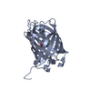
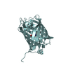
| Deposited unit | 
| ||||||||
|---|---|---|---|---|---|---|---|---|---|
| 1 | 
| ||||||||
| 2 | 
| ||||||||
| 3 | 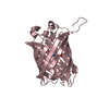
| ||||||||
| 4 | 
| ||||||||
| 5 |
| ||||||||
| Unit cell |
|
-Components
| #1: Protein | Mass: 26247.992 Da / Num. of mol.: 4 / Mutation: S158A Source method: isolated from a genetically manipulated source Source: (gene. exp.) Artificial gene (others) / Production host:  Escherichia coli (E. coli) Escherichia coli (E. coli)#2: Water | ChemComp-HOH / | Water Mass: 18.015 Da / Num. of mol.: 815 / Source method: isolated from a natural source / Formula: H2O Mass: 18.015 Da / Num. of mol.: 815 / Source method: isolated from a natural source / Formula: H2OSequence details | THIS SEQUENCE WAS ENGINEERED USING THE PROTEIN SEQUENCE FP480 FROM ENTACMAEA QUADRICOLOR (UNP ...THIS SEQUENCE WAS ENGINEERED | |
|---|
-Experimental details
-Experiment
| Experiment | Method: X-RAY DIFFRACTION / Number of used crystals: 1 |
|---|
- Sample preparation
| Crystal | Density Matthews: 2.36 Å3/Da / Density % sol: 47.9 % |
|---|---|
| Crystal grow | Temperature: 298.15 K / Method: vapor diffusion, hanging drop / pH: 7.5 Details: 0.1M Tris-HCl, 0.25M ammonium citrate dibasic, 22% PEG3350, pH 7.5, VAPOR DIFFUSION, HANGING DROP, temperature 298.15K |
-Data collection
| Diffraction | Mean temperature: 140 K |
|---|---|
| Diffraction source | Source: SYNCHROTRON / Site: CHESS  / Beamline: A1 / Wavelength: 0.978 Å / Beamline: A1 / Wavelength: 0.978 Å |
| Detector | Type: ADSC QUANTUM 210 / Detector: CCD / Date: Feb 11, 2010 |
| Radiation | Monochromator: GRAPHITE / Protocol: SINGLE WAVELENGTH / Monochromatic (M) / Laue (L): M / Scattering type: x-ray |
| Radiation wavelength | Wavelength: 0.978 Å / Relative weight: 1 |
| Reflection | Resolution: 1.9→50 Å / % possible obs: 100 % / Observed criterion σ(F): 2 / Observed criterion σ(I): 2 |
| Reflection shell | Resolution: 1.9→1.99 Å / % possible all: 100 |
- Processing
| Software |
| ||||||||||||||||||||||||||||||||||||||||||||||||||||||||||||||||||||||||||||||||||||||||||||||||||||||||||||||||||||||||||||||||||||||||||||||||||||||||||||||||||||||||||||||||||||||||||||||||||||
|---|---|---|---|---|---|---|---|---|---|---|---|---|---|---|---|---|---|---|---|---|---|---|---|---|---|---|---|---|---|---|---|---|---|---|---|---|---|---|---|---|---|---|---|---|---|---|---|---|---|---|---|---|---|---|---|---|---|---|---|---|---|---|---|---|---|---|---|---|---|---|---|---|---|---|---|---|---|---|---|---|---|---|---|---|---|---|---|---|---|---|---|---|---|---|---|---|---|---|---|---|---|---|---|---|---|---|---|---|---|---|---|---|---|---|---|---|---|---|---|---|---|---|---|---|---|---|---|---|---|---|---|---|---|---|---|---|---|---|---|---|---|---|---|---|---|---|---|---|---|---|---|---|---|---|---|---|---|---|---|---|---|---|---|---|---|---|---|---|---|---|---|---|---|---|---|---|---|---|---|---|---|---|---|---|---|---|---|---|---|---|---|---|---|---|---|---|---|
| Refinement | Method to determine structure: MOLECULAR REPLACEMENT / Resolution: 1.899→43.933 Å / SU ML: 0.56 / σ(F): 1.34 / Phase error: 22.36 / Stereochemistry target values: ML
| ||||||||||||||||||||||||||||||||||||||||||||||||||||||||||||||||||||||||||||||||||||||||||||||||||||||||||||||||||||||||||||||||||||||||||||||||||||||||||||||||||||||||||||||||||||||||||||||||||||
| Solvent computation | Shrinkage radii: 0.83 Å / VDW probe radii: 1.1 Å / Solvent model: FLAT BULK SOLVENT MODEL / Bsol: 49.985 Å2 / ksol: 0.4 e/Å3 | ||||||||||||||||||||||||||||||||||||||||||||||||||||||||||||||||||||||||||||||||||||||||||||||||||||||||||||||||||||||||||||||||||||||||||||||||||||||||||||||||||||||||||||||||||||||||||||||||||||
| Displacement parameters |
| ||||||||||||||||||||||||||||||||||||||||||||||||||||||||||||||||||||||||||||||||||||||||||||||||||||||||||||||||||||||||||||||||||||||||||||||||||||||||||||||||||||||||||||||||||||||||||||||||||||
| Refinement step | Cycle: LAST / Resolution: 1.899→43.933 Å
| ||||||||||||||||||||||||||||||||||||||||||||||||||||||||||||||||||||||||||||||||||||||||||||||||||||||||||||||||||||||||||||||||||||||||||||||||||||||||||||||||||||||||||||||||||||||||||||||||||||
| Refine LS restraints |
| ||||||||||||||||||||||||||||||||||||||||||||||||||||||||||||||||||||||||||||||||||||||||||||||||||||||||||||||||||||||||||||||||||||||||||||||||||||||||||||||||||||||||||||||||||||||||||||||||||||
| LS refinement shell |
|
