 Movie
Movie Controller

[English] 日本語
 Yorodumi
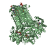
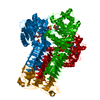
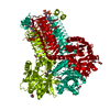
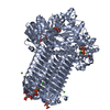
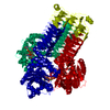
- PDB-3st8: Crystal structure of GlmU from Mycobacterium tuberculosis in comp... -
+ Open data

- Basic information
| Entry | Database: PDB / ID: 3st8 | |||||||||
|---|---|---|---|---|---|---|---|---|---|---|
| Title | Crystal structure of GlmU from Mycobacterium tuberculosis in complex with COENZYME A, GLUCOSAMINE 1-PHOSPHATE and URIDINE-DIPHOSPHATE-N-ACETYLGLUCOSAMINE | |||||||||
 Components Components | Bifunctional protein glmU | |||||||||
 Keywords Keywords | TRANSFERASE / ACETYLTRANSFERASE / BIFUNCTIONAL / PYROPHOSPHORYLASE / ROSSMANN-LIKE FOLD / LEFT-HANDED-BETA-HELIX / CELL SHAPE / CELL WALL BIOGENESIS/DEGRADATION / METAL-BINDING / MULTIFUNCTIONAL ENZYME / NUCLEOTIDYLTRANSFERASE / PEPTIDOGLYCAN SYNTHESIS / ACYLTRANSFERASE | |||||||||
| Function / homology |  Function and homology informationuridylyltransferase activity / adhesion of symbiont to host cell / glucosamine-1-phosphate N-acetyltransferase / glucosamine-1-phosphate N-acetyltransferase activity / UDP-N-acetylglucosamine diphosphorylase / UDP-N-acetylglucosamine diphosphorylase activity / entry of bacterium into host cell / UDP-N-acetylglucosamine biosynthetic process / lipid A biosynthetic process / peptidoglycan biosynthetic process ...uridylyltransferase activity / adhesion of symbiont to host cell / glucosamine-1-phosphate N-acetyltransferase / glucosamine-1-phosphate N-acetyltransferase activity / UDP-N-acetylglucosamine diphosphorylase / UDP-N-acetylglucosamine diphosphorylase activity / entry of bacterium into host cell / UDP-N-acetylglucosamine biosynthetic process / lipid A biosynthetic process / peptidoglycan biosynthetic process / cell wall organization / cell morphogenesis / regulation of cell shape / magnesium ion binding / cytoplasm Function and homology informationuridylyltransferase activity / adhesion of symbiont to host cell / glucosamine-1-phosphate N-acetyltransferase / glucosamine-1-phosphate N-acetyltransferase activity / UDP-N-acetylglucosamine diphosphorylase / UDP-N-acetylglucosamine diphosphorylase activity / entry of bacterium into host cell / UDP-N-acetylglucosamine biosynthetic process / lipid A biosynthetic process / peptidoglycan biosynthetic process ...uridylyltransferase activity / adhesion of symbiont to host cell / glucosamine-1-phosphate N-acetyltransferase / glucosamine-1-phosphate N-acetyltransferase activity / UDP-N-acetylglucosamine diphosphorylase / UDP-N-acetylglucosamine diphosphorylase activity / entry of bacterium into host cell / UDP-N-acetylglucosamine biosynthetic process / lipid A biosynthetic process / peptidoglycan biosynthetic process / cell wall organization / cell morphogenesis / regulation of cell shape / magnesium ion binding / cytoplasmSimilarity search - Function | |||||||||
| Biological species |   Mycobacterium tuberculosis (bacteria) Mycobacterium tuberculosis (bacteria) | |||||||||
| Method | X-RAY DIFFRACTION / SYNCHROTRON / MOLECULAR REPLACEMENT / Resolution: 1.98 Å | |||||||||
 Authors Authors | Jagtap, P.A. / Prakash, B. | |||||||||
 Citation Citation | Journal: to be published Title: Structure of Mycobacterium tuberculosis GlmU in complex with Acetyl CoA Authors: Jagtap, P.A. / Prakash, B. | |||||||||
| History |
|
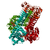
- Structure visualization
| Structure viewer | Molecule: MolmilJmol/JSmol |
|---|
- Downloads & links
-Download
| PDBx/mmCIF format | 3st8.cif.gz | 123 KB | Display | PDBx/mmCIF format |
|---|---|---|---|---|
| PDB format | pdb3st8.ent.gz | 89.7 KB | Display | PDB format |
| PDBx/mmJSON format | 3st8.json.gz | Tree view | PDBx/mmJSON format | |
| Others |  Other downloads Other downloads |
-Validation report
| Arichive directory | https://data.pdbj.org/pub/pdb/validation_reports/st/3st8ftp://data.pdbj.org/pub/pdb/validation_reports/st/3st8 | HTTPS FTP |
|---|
-Related structure data
| Related structure data |  3sptC  3spm 3spn 3spo 3spp 3spq C: citing same article ( |
|---|---|
| Similar structure data |
-Links
 PDBj
PDBj


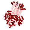
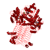
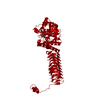
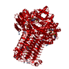
- Assembly
| Deposited unit | 
| |||||||||||||||||||||||||||
|---|---|---|---|---|---|---|---|---|---|---|---|---|---|---|---|---|---|---|---|---|---|---|---|---|---|---|---|---|
| 1 | 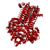
| |||||||||||||||||||||||||||
| Unit cell |
| |||||||||||||||||||||||||||
| Components on special symmetry positions |
|
-Components
-Protein / Sugars , 2 types, 2 molecules A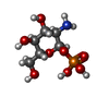
| #1: Protein | / UDP-N-acetylglucosamine pyrophosphorylase / N-acetylglucosamine-1-phosphate uridyltransferase / ...UDP-N-acetylglucosamine pyrophosphorylase / N-acetylglucosamine-1-phosphate uridyltransferase / Glucosamine-1-phosphate N-acetyltransferase Mass: 52466.602 Da / Num. of mol.: 1 Source method: isolated from a genetically manipulated source Source: (gene. exp.) Mycobacterium tuberculosis (bacteria) / Strain: H37Rv / Gene: glmU, MT1046, Rv1018c / Plasmid: pQEII / Production host: Escherichia coli (E. coli) / Strain (production host): DH5ALPHAReferences: UniProt: P96382, UniProt: P9WMN3*PLUS, UDP-N-acetylglucosamine diphosphorylase, glucosamine-1-phosphate N-acetyltransferase |
|---|---|
| #4: Sugar | ChemComp-GP1 /  Type: D-saccharide / Mass: 259.151 Da / Num. of mol.: 1 Type: D-saccharide / Mass: 259.151 Da / Num. of mol.: 1Source method: isolated from a genetically manipulated source Formula: C6H14NO8P |
-Non-polymers , 4 types, 535 molecules 


| #2: Chemical | ChemComp-MG /  Mass: 24.305 Da / Num. of mol.: 4 / Source method: obtained synthetically / Formula: Mg Mass: 24.305 Da / Num. of mol.: 4 / Source method: obtained synthetically / Formula: Mg#3: Chemical | ChemComp-COA / | Coenzyme A Mass: 767.534 Da / Num. of mol.: 1 / Source method: obtained synthetically / Formula: C21H36N7O16P3S Mass: 767.534 Da / Num. of mol.: 1 / Source method: obtained synthetically / Formula: C21H36N7O16P3S#5: Chemical | ChemComp-UD1 / |  Mass: 607.354 Da / Num. of mol.: 1 / Source method: obtained synthetically / Formula: C17H27N3O17P2 Mass: 607.354 Da / Num. of mol.: 1 / Source method: obtained synthetically / Formula: C17H27N3O17P2#6: Water | ChemComp-HOH / | WaterMass: 18.015 Da / Num. of mol.: 529 / Source method: isolated from a natural source / Formula: H2O |
|---|
-Experimental details
-Experiment
| Experiment | Method: X-RAY DIFFRACTION / Number of used crystals: 1 |
|---|
- Sample preparation
| Crystal | Density Matthews: 4.02 Å3/Da / Density % sol: 69.43 % |
|---|---|
| Crystal grow | Temperature: 291 K / Method: vapor diffusion, sitting drop / pH: 8.5 Details: 0.1M Tris-Cl, pH-8.5, 2% Tacsimate, 18% PEG 3350, VAPOR DIFFUSION, SITTING DROP, temperature 291K |
-Data collection
| Diffraction | Mean temperature: 100 K | ||||||||||||||||||||||||||||||||||||||||||||||||||||||||||||||||||||||||||||||||||||||||||||||||||||||||||||||||||||||||||||||||||||||||||||||||||||||||||||||||||||||||||||||||||||||||||||||||||||||||||||||||||
|---|---|---|---|---|---|---|---|---|---|---|---|---|---|---|---|---|---|---|---|---|---|---|---|---|---|---|---|---|---|---|---|---|---|---|---|---|---|---|---|---|---|---|---|---|---|---|---|---|---|---|---|---|---|---|---|---|---|---|---|---|---|---|---|---|---|---|---|---|---|---|---|---|---|---|---|---|---|---|---|---|---|---|---|---|---|---|---|---|---|---|---|---|---|---|---|---|---|---|---|---|---|---|---|---|---|---|---|---|---|---|---|---|---|---|---|---|---|---|---|---|---|---|---|---|---|---|---|---|---|---|---|---|---|---|---|---|---|---|---|---|---|---|---|---|---|---|---|---|---|---|---|---|---|---|---|---|---|---|---|---|---|---|---|---|---|---|---|---|---|---|---|---|---|---|---|---|---|---|---|---|---|---|---|---|---|---|---|---|---|---|---|---|---|---|---|---|---|---|---|---|---|---|---|---|---|---|---|---|---|---|---|
| Diffraction source | Source: SYNCHROTRON / Site: ESRF  / Beamline: BM14 / Wavelength: 0.97625 Å / Beamline: BM14 / Wavelength: 0.97625 Å | ||||||||||||||||||||||||||||||||||||||||||||||||||||||||||||||||||||||||||||||||||||||||||||||||||||||||||||||||||||||||||||||||||||||||||||||||||||||||||||||||||||||||||||||||||||||||||||||||||||||||||||||||||
| Detector | Type: MARCCD 225 / Detector: CCD / Date: Jan 29, 2011 / Details: mirrors | ||||||||||||||||||||||||||||||||||||||||||||||||||||||||||||||||||||||||||||||||||||||||||||||||||||||||||||||||||||||||||||||||||||||||||||||||||||||||||||||||||||||||||||||||||||||||||||||||||||||||||||||||||
| Radiation | Protocol: SINGLE WAVELENGTH / Monochromatic (M) / Laue (L): M / Scattering type: x-ray | ||||||||||||||||||||||||||||||||||||||||||||||||||||||||||||||||||||||||||||||||||||||||||||||||||||||||||||||||||||||||||||||||||||||||||||||||||||||||||||||||||||||||||||||||||||||||||||||||||||||||||||||||||
| Radiation wavelength | Wavelength: 0.97625 Å / Relative weight: 1 | ||||||||||||||||||||||||||||||||||||||||||||||||||||||||||||||||||||||||||||||||||||||||||||||||||||||||||||||||||||||||||||||||||||||||||||||||||||||||||||||||||||||||||||||||||||||||||||||||||||||||||||||||||
| Reflection | Number: 432608 / Rmerge(I) obs: 0.053 / D res high: 1.98 Å / Num. obs: 58849 / % possible obs: 99.8 | ||||||||||||||||||||||||||||||||||||||||||||||||||||||||||||||||||||||||||||||||||||||||||||||||||||||||||||||||||||||||||||||||||||||||||||||||||||||||||||||||||||||||||||||||||||||||||||||||||||||||||||||||||
| Diffraction reflection shell |
| ||||||||||||||||||||||||||||||||||||||||||||||||||||||||||||||||||||||||||||||||||||||||||||||||||||||||||||||||||||||||||||||||||||||||||||||||||||||||||||||||||||||||||||||||||||||||||||||||||||||||||||||||||
| Reflection | Resolution: 1.98→19.71 Å / Num. all: 58954 / Num. obs: 58849 / % possible obs: 99.8 % / Observed criterion σ(F): 0 / Observed criterion σ(I): -3 / Redundancy: 7.3 % / Biso Wilson estimate: 30.049 Å2 / Rmerge(I) obs: 0.053 / Rsym value: 0.057 / Net I/σ(I): 25.88 | ||||||||||||||||||||||||||||||||||||||||||||||||||||||||||||||||||||||||||||||||||||||||||||||||||||||||||||||||||||||||||||||||||||||||||||||||||||||||||||||||||||||||||||||||||||||||||||||||||||||||||||||||||
| Reflection shell | Diffraction-ID: 1
|
- Processing
| Software |
| |||||||||||||||||||||||||||||||||||||||||||||||||||||||||||||||||
|---|---|---|---|---|---|---|---|---|---|---|---|---|---|---|---|---|---|---|---|---|---|---|---|---|---|---|---|---|---|---|---|---|---|---|---|---|---|---|---|---|---|---|---|---|---|---|---|---|---|---|---|---|---|---|---|---|---|---|---|---|---|---|---|---|---|---|
| Refinement | Method to determine structure: MOLECULAR REPLACEMENT / Resolution: 1.98→19.71 Å / Cor.coef. Fo:Fc: 0.96 / Cor.coef. Fo:Fc free: 0.943 / WRfactor Rfree: 0.1945 / WRfactor Rwork: 0.1647 / Occupancy max: 1 / Occupancy min: 0.33 / FOM work R set: 0.8829 / SU B: 2.363 / SU ML: 0.068 / SU R Cruickshank DPI: 0.1056 / SU Rfree: 0.1076 / Cross valid method: THROUGHOUT / σ(F): 0 / ESU R Free: 0.108 / Stereochemistry target values: MAXIMUM LIKELIHOOD Details: HYDROGENS HAVE BEEN ADDED IN THE RIDING POSITIONS U VALUES : REFINED INDIVIDUALLY
| |||||||||||||||||||||||||||||||||||||||||||||||||||||||||||||||||
| Solvent computation | Ion probe radii: 0.8 Å / Shrinkage radii: 0.8 Å / VDW probe radii: 1.4 Å / Solvent model: MASK | |||||||||||||||||||||||||||||||||||||||||||||||||||||||||||||||||
| Displacement parameters | Biso max: 500 Å2 / Biso mean: 26.3707 Å2 / Biso min: 6.16 Å2
| |||||||||||||||||||||||||||||||||||||||||||||||||||||||||||||||||
| Refinement step | Cycle: LAST / Resolution: 1.98→19.71 Å
| |||||||||||||||||||||||||||||||||||||||||||||||||||||||||||||||||
| Refine LS restraints |
| |||||||||||||||||||||||||||||||||||||||||||||||||||||||||||||||||
| LS refinement shell | Resolution: 1.984→2.035 Å / Total num. of bins used: 20
|
