 Movie
Movie Controller

[English] 日本語
 Yorodumi
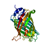
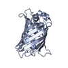
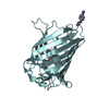
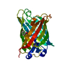
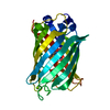
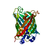
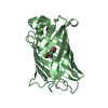
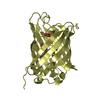
- PDB-3ssv: Engineered low-affinity halide-binding protein derived from YFP: ... -
+ Open data

- Basic information
| Entry | Database: PDB / ID: 3ssv | |||||||||
|---|---|---|---|---|---|---|---|---|---|---|
| Title | Engineered low-affinity halide-binding protein derived from YFP: fluoride complex | |||||||||
 Components Components | Green fluorescent protein | |||||||||
 Keywords Keywords | HALIDE BINDING PROTEIN / beta barrel / luminescent protein / yellow fluorescent protein / imaging reagent | |||||||||
| Function / homology |  Function and homology information Function and homology informationserine-type endopeptidase inhibitor activity / extracellular space / metal ion binding Similarity search - Function | |||||||||
| Biological species |   Aequorea victoria (jellyfish) Aequorea victoria (jellyfish) | |||||||||
| Method |  X-RAY DIFFRACTION / SYNCHROTRON / FOURIER SYNTHESIS / Resolution: 1.861 Å X-RAY DIFFRACTION / SYNCHROTRON / FOURIER SYNTHESIS / Resolution: 1.861 Å | |||||||||
 Authors Authors | Wang, W. / Grimley, J.S. / Beese, L.S. / Hellinga, H.W. | |||||||||
 Citation Citation | Journal: To be Published Title: Determination of engineered chloride-binding site structures in fluorescent proteins reveals principles of halide recognition Authors: Wang, W. / Grimley, J.S. / Augustine, G.J. / Beese, L.S. / Hellinga, H.W. | |||||||||
| History |
|
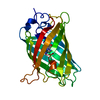
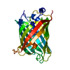
- Structure visualization
| Structure viewer | Molecule: MolmilJmol/JSmol |
|---|
- Downloads & links
-Download
| PDBx/mmCIF format | 3ssv.cif.gz | 104.3 KB | Display | PDBx/mmCIF format |
|---|---|---|---|---|
| PDB format | pdb3ssv.ent.gz | 78.8 KB | Display | PDB format |
| PDBx/mmJSON format | 3ssv.json.gz | Tree view | PDBx/mmJSON format | |
| Others |  Other downloads Other downloads |
-Validation report
| Summary document | 3ssv_validation.pdf.gz | 422.5 KB | Display | wwPDB validaton report |
|---|---|---|---|---|
| Full document | 3ssv_full_validation.pdf.gz | 424.2 KB | Display | |
| Data in XML | 3ssv_validation.xml.gz | 12.8 KB | Display | |
| Data in CIF | 3ssv_validation.cif.gz | 18.1 KB | Display | |
| Arichive directory | https://data.pdbj.org/pub/pdb/validation_reports/ss/3ssvftp://data.pdbj.org/pub/pdb/validation_reports/ss/3ssv | HTTPS FTP |
-Related structure data
| Related structure data | 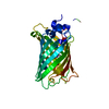 3sryC 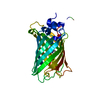 3ss0C 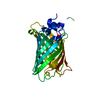 3sshC  3sskC  3sslC 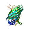 3sspC 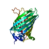 3sstSC 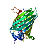 3ssyC 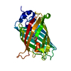 3svbC 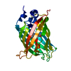 3svcC  3svdC 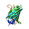 3sveC C: citing same article ( S: Starting model for refinement |
|---|---|
| Similar structure data |


-Links
 PDBj
PDBj


- Assembly
| Deposited unit | 
| ||||||||
|---|---|---|---|---|---|---|---|---|---|
| 1 |
| ||||||||
| Unit cell |
|
-Components
| #1: Protein | Mass: 29362.801 Da / Num. of mol.: 1 / Mutation: S72A, K79R, T203Y, H231L Source method: isolated from a genetically manipulated source Details: synthetic / Source: (gene. exp.) Aequorea victoria (jellyfish) / Gene: GFP / Plasmid: pUC19 / Production host:  |
|---|---|
| #2: Chemical | ChemComp-F /   Mass: 18.998 Da / Num. of mol.: 1 / Source method: obtained synthetically / Formula: F Mass: 18.998 Da / Num. of mol.: 1 / Source method: obtained synthetically / Formula: F |
| #3: Water | ChemComp-HOH /  Mass: 18.015 Da / Num. of mol.: 193 / Source method: isolated from a natural source / Formula: H2O Mass: 18.015 Da / Num. of mol.: 193 / Source method: isolated from a natural source / Formula: H2O |
| Sequence details | UNP RESIDUE SER65 UNDERWENT MUTATION TO GLY. GLY65, TYR66, AND GLY67 CIRCULARIZ |
-Experimental details
-Experiment
| Experiment | Method: X-RAY DIFFRACTION / Number of used crystals: 1 |
|---|
- Sample preparation
| Crystal | Density Matthews: 1.92 Å3/Da / Density % sol: 35.96 % |
|---|---|
| Crystal grow | Temperature: 290 K / Method: vapor diffusion, hanging drop / pH: 5.6 Details: 18% PEG2000, 50 mM sodium acetate, 90 mM magnesium chloride, pH 5.6, VAPOR DIFFUSION, HANGING DROP, temperature 290K |
-Data collection
| Diffraction | Mean temperature: 100 K |
|---|---|
| Diffraction source | Source: SYNCHROTRON / Site: APS  / Beamline: 22-ID / Wavelength: 1 Å / Beamline: 22-ID / Wavelength: 1 Å |
| Detector | Type: MARMOSAIC 300 mm CCD / Detector: CCD / Date: Dec 17, 2009 |
| Radiation | Monochromator: Rosenbaum-Rock double-crystal Si(111) / Protocol: SINGLE WAVELENGTH / Monochromatic (M) / Laue (L): M / Scattering type: x-ray |
| Radiation wavelength | Wavelength: 1 Å / Relative weight: 1 |
| Reflection | Resolution: 1.861→46.798 Å / Num. all: 19658 / Num. obs: 19414 |
- Processing
| Software |
| ||||||||||||||||||||||||||||||||||||||||||||||||||||||||
|---|---|---|---|---|---|---|---|---|---|---|---|---|---|---|---|---|---|---|---|---|---|---|---|---|---|---|---|---|---|---|---|---|---|---|---|---|---|---|---|---|---|---|---|---|---|---|---|---|---|---|---|---|---|---|---|---|---|
| Refinement | Method to determine structure: FOURIER SYNTHESIS Starting model: PDB ENTRY 3SST Resolution: 1.861→46.798 Å / Occupancy max: 1 / Occupancy min: 0 / SU ML: 0.41 / σ(F): 1.34 / Phase error: 21.52 / Stereochemistry target values: ML
| ||||||||||||||||||||||||||||||||||||||||||||||||||||||||
| Solvent computation | Shrinkage radii: 0.77 Å / VDW probe radii: 0.9 Å / Solvent model: FLAT BULK SOLVENT MODEL / Bsol: 38.977 Å2 / ksol: 0.402 e/Å3 | ||||||||||||||||||||||||||||||||||||||||||||||||||||||||
| Displacement parameters |
| ||||||||||||||||||||||||||||||||||||||||||||||||||||||||
| Refinement step | Cycle: LAST / Resolution: 1.861→46.798 Å
| ||||||||||||||||||||||||||||||||||||||||||||||||||||||||
| Refine LS restraints |
| ||||||||||||||||||||||||||||||||||||||||||||||||||||||||
| LS refinement shell |
|
