 Movie
Movie Controller


+ Open data

- Basic information
| Entry | Database: PDB / ID: 3spa | ||||||
|---|---|---|---|---|---|---|---|
| Title | Crystal Structure of Human Mitochondrial RNA Polymerase | ||||||
 Components Components |
| ||||||
 Keywords Keywords |  TRANSFERASE / single-subunit DNA-dependent RNA Polymerase in mitochondria / Mitochondria TRANSFERASE / single-subunit DNA-dependent RNA Polymerase in mitochondria / Mitochondria | ||||||
| Function / homology |  Function and homology information Function and homology informationMitochondrial transcription initiation / mitochondrial DNA-directed RNA polymerase complex / mitochondrial promoter sequence-specific DNA binding / transcription initiation at mitochondrial promoter / mitochondrial transcription / DNA primase activity / mitochondrial nucleoid / Transcriptional activation of mitochondrial biogenesis / DNA-directed 5'-3' RNA polymerase activity / DNA-directed RNA polymerase ...Mitochondrial transcription initiation / mitochondrial DNA-directed RNA polymerase complex / mitochondrial promoter sequence-specific DNA binding / transcription initiation at mitochondrial promoter / mitochondrial transcription / DNA primase activity / mitochondrial nucleoid / Transcriptional activation of mitochondrial biogenesis / DNA-directed 5'-3' RNA polymerase activity / DNA-directed RNA polymerase / 3'-5'-RNA exonuclease activity / sequence-specific DNA binding / mitochondrial matrix / protein-containing complex / mitochondrion / RNA bindingSimilarity search - Function | ||||||
| Biological species |  Homo sapiens (human) Homo sapiens (human) | ||||||
| Method | X-RAY DIFFRACTION / SYNCHROTRON / MOLECULAR REPLACEMENT / Resolution: 2.5 Å | ||||||
 Authors Authors | Ringel, R. / Sologub, M. / Morozov, Y.I. / Litonin, D. / Cramer, P. / Temiakov, D. | ||||||
 Citation Citation | Journal: Nature / Year: 2011 Title: Structure of human mitochondrial RNA polymerase Authors: Ringel, R. / Sologub, M. / Morozov, Y.I. / Litonin, D. / Cramer, P. / Temiakov, D. | ||||||
| History |
| ||||||
| Remark 999 | THE AUTHOR STATES THAT CHAINS B IS LIKELY PARTS OF THE N-TERMINI OF CHAINS A. DUD TO DISORDER, IT ...THE AUTHOR STATES THAT CHAINS B IS LIKELY PARTS OF THE N-TERMINI OF CHAINS A. DUD TO DISORDER, IT WAS NOT POSSIBLE TO ACCURATELY ASSIGN THE SIDE CHAINS AND IDENTIFY THE PEPTIDES. SO THEY ARE MODELED AS UNK. |
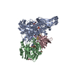
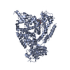
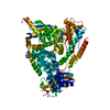
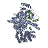
- Structure visualization
| Structure viewer | Molecule: MolmilJmol/JSmol |
|---|
- Downloads & links
-Download
| PDBx/mmCIF format | 3spa.cif.gz | 395.5 KB | Display | PDBx/mmCIF format |
|---|---|---|---|---|
| PDB format | pdb3spa.ent.gz | 320.5 KB | Display | PDB format |
| PDBx/mmJSON format | 3spa.json.gz | Tree view | PDBx/mmJSON format | |
| Others |  Other downloads Other downloads |
-Validation report
| Arichive directory | https://data.pdbj.org/pub/pdb/validation_reports/sp/3spaftp://data.pdbj.org/pub/pdb/validation_reports/sp/3spa | HTTPS FTP |
|---|
-Related structure data
| Related structure data |  1qlnS S: Starting model for refinement |
|---|---|
| Similar structure data |


-Links
 PDBj
PDBj
- Assembly
| Deposited unit | 
| ||||||||
|---|---|---|---|---|---|---|---|---|---|
| 1 |
| ||||||||
| Unit cell |
|
-Components
-Protein / Protein/peptide , 2 types, 2 molecules AB
| #1: Protein | Polymerase / MtRPOL Mass: 128736.031 Da / Num. of mol.: 1 Source method: isolated from a genetically manipulated source Source: (gene. exp.) Homo sapiens (human) / Gene: AAC06147.1, POLRMT / Plasmid: pProExHtb / Production host:  Escherichia coli (E. coli) / References: UniProt: O00411, DNA-directed RNA polymerase Escherichia coli (E. coli) / References: UniProt: O00411, DNA-directed RNA polymerase |
|---|---|
| #2: Protein/peptide | Mass: 783.958 Da / Num. of mol.: 1 Source method: isolated from a genetically manipulated source Source: (gene. exp.) Homo sapiens (human) / Gene: AAC06147.1, POLRMT / Plasmid: pProExHtb / Production host: Escherichia coli (E. coli) |
-Non-polymers , 4 types, 153 molecules 


| #3: Chemical | Glycerol Mass: 92.094 Da / Num. of mol.: 2 / Source method: obtained synthetically / Formula: C3H8O3 Mass: 92.094 Da / Num. of mol.: 2 / Source method: obtained synthetically / Formula: C3H8O3#4: Chemical | ChemComp-CL / Chloride Mass: 35.453 Da / Num. of mol.: 5 / Source method: obtained synthetically / Formula: Cl Mass: 35.453 Da / Num. of mol.: 5 / Source method: obtained synthetically / Formula: Cl#5: Chemical | ChemComp-SO4 / | Sulfate Mass: 96.063 Da / Num. of mol.: 1 / Source method: obtained synthetically / Formula: SO4 Mass: 96.063 Da / Num. of mol.: 1 / Source method: obtained synthetically / Formula: SO4#6: Water | ChemComp-HOH / | WaterMass: 18.015 Da / Num. of mol.: 145 / Source method: isolated from a natural source / Formula: H2O |
|---|
-Experimental details
-Experiment
| Experiment | Method: X-RAY DIFFRACTION / Number of used crystals: 1 |
|---|
- Sample preparation
| Crystal | Density Matthews: 2.6 Å3/Da / Density % sol: 52.77 % |
|---|---|
| Crystal grow | Temperature: 293 K / Method: vapor diffusion, hanging drop / pH: 6.5 Details: 12% PEG4000, 10% Glycerol, 30mM Mes 6.5, 60mM (NH4)2SO4, 40mM Tris 8.0, 20mM DTT in situ proteolysed with ArgC (1:1000 w/w ratio) and micro-seeded, VAPOR DIFFUSION, HANGING DROP, temperature 293K |
-Data collection
| Diffraction | Mean temperature: 63 K |
|---|---|
| Diffraction source | Source: SYNCHROTRON / Site: SLS  / Beamline: X06DA / Wavelength: 1 Å / Beamline: X06DA / Wavelength: 1 Å |
| Detector | Type: MARMOSAIC 225 mm CCD / Detector: CCD / Date: Feb 4, 2011 |
| Radiation | Monochromator: Bartels Monochromator / Protocol: SINGLE WAVELENGTH / Monochromatic (M) / Laue (L): M / Scattering type: x-ray |
| Radiation wavelength | Wavelength: 1 Å / Relative weight: 1 |
| Reflection | Resolution: 2.5→58.128 Å / Num. all: 46595 / Num. obs: 46507 / Redundancy: 3.8 % / Biso Wilson estimate: 73.16 Å2 / Rmerge(I) obs: 0.047 / Net I/σ(I): 15.9 |
| Reflection shell | Resolution: 2.5→2.64 Å / Redundancy: 3.8 % / Rmerge(I) obs: 0.631 / Mean I/σ(I) obs: 1.9 / Num. unique all: 6694 |
- Processing
| Software |
| ||||||||||||||||||||||||||||||||||||||||||||||||||||||||||||||||||
|---|---|---|---|---|---|---|---|---|---|---|---|---|---|---|---|---|---|---|---|---|---|---|---|---|---|---|---|---|---|---|---|---|---|---|---|---|---|---|---|---|---|---|---|---|---|---|---|---|---|---|---|---|---|---|---|---|---|---|---|---|---|---|---|---|---|---|---|
| Refinement | Method to determine structure: MOLECULAR REPLACEMENT Starting model: PDB entry 1QLN, truncated to polyAlanine, residues 246-354, 392-568, 691-741, 766-883 of model used Resolution: 2.5→58.128 Å / Cor.coef. Fo:Fc: 0.9494 / Cor.coef. Fo:Fc free: 0.9259 / SU R Cruickshank DPI: 0.327 / Cross valid method: THROUGHOUT / σ(F): 0 / Stereochemistry target values: Engh & Huber Details: Residues 326-360 were refined with correct sequence and afterwards rebuild to poly Alanine because of poor side chain density and possible sequence shifts in this region. PDB includes these ...Details: Residues 326-360 were refined with correct sequence and afterwards rebuild to poly Alanine because of poor side chain density and possible sequence shifts in this region. PDB includes these residues as UNK. An unconnected helix is build as poly Alanine and also named with UNK residues in the PDB. Residues 1002-1005 possibly have more conformations than built, explaining additional positive density.
| ||||||||||||||||||||||||||||||||||||||||||||||||||||||||||||||||||
| Displacement parameters | Biso mean: 89.21 Å2
| ||||||||||||||||||||||||||||||||||||||||||||||||||||||||||||||||||
| Refine analyze | Luzzati coordinate error obs: 0.387 Å | ||||||||||||||||||||||||||||||||||||||||||||||||||||||||||||||||||
| Refinement step | Cycle: LAST / Resolution: 2.5→58.128 Å
| ||||||||||||||||||||||||||||||||||||||||||||||||||||||||||||||||||
| Refine LS restraints |
| ||||||||||||||||||||||||||||||||||||||||||||||||||||||||||||||||||
| LS refinement shell | Resolution: 2.5→2.56 Å / Total num. of bins used: 20
| ||||||||||||||||||||||||||||||||||||||||||||||||||||||||||||||||||
| Refinement TLS params. | Method: refined / Origin x: 3.3823 Å / Origin y: 41.4659 Å / Origin z: -0.7464 Å
| ||||||||||||||||||||||||||||||||||||||||||||||||||||||||||||||||||
| Refinement TLS group | Selection details: { A|* } |
