 Movie
Movie Controller

[English] 日本語
 Yorodumi
- PDB-3soo: Crystal structure of a LINE-1 type transposase domain-containing ... -
+ Open data

- Basic information
| Entry | Database: PDB / ID: 3soo | ||||||
|---|---|---|---|---|---|---|---|
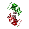
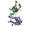
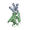
| Title | Crystal structure of a LINE-1 type transposase domain-containing protein 1 (L1TD1) from HOMO SAPIENS at 2.73 A resolution | ||||||
 Components Components | LINE-1 type transposase domain-containing protein 1 | ||||||
 Keywords Keywords |  RNA BINDING PROTEIN / Alpha/beta protein / Structural Genomics / Joint Center for Structural Genomics / JCSG / Protein Structure Initiative / PSI-BIOLOGY / Partnership for Stem Cell Biology / STEMCELL RNA BINDING PROTEIN / Alpha/beta protein / Structural Genomics / Joint Center for Structural Genomics / JCSG / Protein Structure Initiative / PSI-BIOLOGY / Partnership for Stem Cell Biology / STEMCELL | ||||||
| Function / homology |  Function and homology informationretrotransposition / single-stranded RNA binding / ribonucleoprotein complex / intracellular membrane-bounded organelle Function and homology informationretrotransposition / single-stranded RNA binding / ribonucleoprotein complex / intracellular membrane-bounded organelleSimilarity search - Function | ||||||
| Biological species |  Homo sapiens (human) Homo sapiens (human) | ||||||
| Method | X-RAY DIFFRACTION / SYNCHROTRON / MAD / Resolution: 2.73 Å | ||||||
 Authors Authors | Joint Center for Structural Genomics (JCSG) / Partnership for Stem Cell Biology (STEMCELL) | ||||||
 Citation Citation | Journal: To be published Title: Crystal structure of a LINE-1 type transposase domain-containing protein 1 (L1TD1) from HOMO SAPIENS at 2.73 A resolution Authors: Joint Center for Structural Genomics (JCSG) / Partnership for Stem Cell Biology (STEMCELL) | ||||||
| History |
|
- Structure visualization
| Structure viewer | Molecule: MolmilJmol/JSmol |
|---|
- Downloads & links
-Download
| PDBx/mmCIF format | 3soo.cif.gz | 114 KB | Display | PDBx/mmCIF format |
|---|---|---|---|---|
| PDB format | pdb3soo.ent.gz | 95.4 KB | Display | PDB format |
| PDBx/mmJSON format | 3soo.json.gz | Tree view | PDBx/mmJSON format | |
| Others |  Other downloads Other downloads |
-Validation report
| Arichive directory | https://data.pdbj.org/pub/pdb/validation_reports/so/3sooftp://data.pdbj.org/pub/pdb/validation_reports/so/3soo | HTTPS FTP |
|---|
-Related structure data
| Similar structure data | |
|---|---|
| Other databases |


-Links
 PDBj
PDBj- Assembly
| Deposited unit | 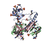
| ||||||||
|---|---|---|---|---|---|---|---|---|---|
| 1 | 
| ||||||||
| 2 | 
| ||||||||
| Unit cell |
| ||||||||
| Components on special symmetry positions |
|
-Components
| #1: Protein | Mass: 10275.656 Da / Num. of mol.: 3 / Fragment: residues 235-321 Source method: isolated from a genetically manipulated source Source: (gene. exp.) Homo sapiens (human) / Gene: BC111559, ECAT11, L1TD1 / Plasmid: SpeedET / Production host:  Escherichia Coli (E. coli) / Strain (production host): HK100 / References: UniProt: Q5T7N2 Escherichia Coli (E. coli) / Strain (production host): HK100 / References: UniProt: Q5T7N2#2: Chemical | ChemComp-SO4 / Sulfate  Mass: 96.063 Da / Num. of mol.: 8 / Source method: obtained synthetically / Formula: SO4 Mass: 96.063 Da / Num. of mol.: 8 / Source method: obtained synthetically / Formula: SO4#3: Chemical | ChemComp-CL / | Chloride  Mass: 35.453 Da / Num. of mol.: 1 / Source method: obtained synthetically / Formula: Cl Mass: 35.453 Da / Num. of mol.: 1 / Source method: obtained synthetically / Formula: ClSequence details | 1. THE CONSTRUCT (RESIDUES 235-321) WAS EXPRESSED WITH A N-TERMINAL PURIFICATION TAG ...1. THE CONSTRUCT (RESIDUES 235-321) WAS EXPRESSED WITH A N-TERMINAL PURIFICATI | |
|---|
-Experimental details
-Experiment
| Experiment | Method: X-RAY DIFFRACTION / Number of used crystals: 1 |
|---|
- Sample preparation
| Crystal | Density Matthews: 3.27 Å3/Da / Density % sol: 62.39 % Description: While data were scaled with XSCALE, the final statistics reported in the REMARK 200 section have been calculated with SCALA with fixed scales and no sd correction. |
|---|---|
| Crystal grow | Temperature: 277 K / Method: vapor diffusion, sitting drop / pH: 5.6 Details: 0.2M potassium sodium tartrate, 2.0M ammonium sulfate, 0.1M sodium citrate pH 5.6, NANODROP', VAPOR DIFFUSION, SITTING DROP, temperature 277K |
-Data collection
| Diffraction | Mean temperature: 100 K | ||||||||||||||||||||||||||||||||||||||||||||||||||||||||||||||||||||||||||||||||||||||||
|---|---|---|---|---|---|---|---|---|---|---|---|---|---|---|---|---|---|---|---|---|---|---|---|---|---|---|---|---|---|---|---|---|---|---|---|---|---|---|---|---|---|---|---|---|---|---|---|---|---|---|---|---|---|---|---|---|---|---|---|---|---|---|---|---|---|---|---|---|---|---|---|---|---|---|---|---|---|---|---|---|---|---|---|---|---|---|---|---|---|
| Diffraction source | Source: SYNCHROTRON / Site: SSRL  / Beamline: BL9-2 / Wavelength: 0.91162,0.97932,0.97907 / Beamline: BL9-2 / Wavelength: 0.91162,0.97932,0.97907 | ||||||||||||||||||||||||||||||||||||||||||||||||||||||||||||||||||||||||||||||||||||||||
| Detector | Type: MARMOSAIC 325 mm CCD / Detector: CCD / Date: Apr 21, 2011 / Details: double crystal monochromator | ||||||||||||||||||||||||||||||||||||||||||||||||||||||||||||||||||||||||||||||||||||||||
| Radiation | Monochromator: double crystal / Protocol: MAD / Monochromatic (M) / Laue (L): M / Scattering type: x-ray | ||||||||||||||||||||||||||||||||||||||||||||||||||||||||||||||||||||||||||||||||||||||||
| Radiation wavelength |
| ||||||||||||||||||||||||||||||||||||||||||||||||||||||||||||||||||||||||||||||||||||||||
| Reflection | Resolution: 2.73→48.712 Å / Num. all: 11913 / Num. obs: 11913 / % possible obs: 99.8 % / Redundancy: 6.7 % / Rsym value: 0.063 / Net I/σ(I): 15.2 | ||||||||||||||||||||||||||||||||||||||||||||||||||||||||||||||||||||||||||||||||||||||||
| Reflection shell | Diffraction-ID: 1
|
-Phasing
| Phasing | Method: MAD |
|---|
- Processing
| Software |
| ||||||||||||||||||||||||||||||||||||||||||||||||||||||||||||||||||||||||||||||||||||||||||||||||||||||||||||
|---|---|---|---|---|---|---|---|---|---|---|---|---|---|---|---|---|---|---|---|---|---|---|---|---|---|---|---|---|---|---|---|---|---|---|---|---|---|---|---|---|---|---|---|---|---|---|---|---|---|---|---|---|---|---|---|---|---|---|---|---|---|---|---|---|---|---|---|---|---|---|---|---|---|---|---|---|---|---|---|---|---|---|---|---|---|---|---|---|---|---|---|---|---|---|---|---|---|---|---|---|---|---|---|---|---|---|---|---|---|
| Refinement | Method to determine structure: MAD / Resolution: 2.73→48.712 Å / Cor.coef. Fo:Fc: 0.931 / Cor.coef. Fo:Fc free: 0.9186 / Occupancy max: 1 / Occupancy min: 0.5 / Cross valid method: THROUGHOUT / σ(F): 0 Details: 1. A MET-INHIBITION PROTOCOL WAS USED FOR SELENOMETHIONINE INCORPORATION DURING PROTEIN EXPRESSION. THE OCCUPANCY OF THE SE ATOMS IN THE MSE RESIDUES WAS REDUCED TO 0.75 FOR THE REDUCED ...Details: 1. A MET-INHIBITION PROTOCOL WAS USED FOR SELENOMETHIONINE INCORPORATION DURING PROTEIN EXPRESSION. THE OCCUPANCY OF THE SE ATOMS IN THE MSE RESIDUES WAS REDUCED TO 0.75 FOR THE REDUCED SCATTERING POWER DUE TO PARTIAL S-MET INCORPORATION. 2. ATOM RECORD CONTAINS SUM OF TLS AND RESIDUAL B FACTORS. ANISOU RECORD CONTAINS SUM OF TLS AND RESIDUAL U FACTORS. 3. SULFATE, CHLORIDE MODELED ARE PRESENT IN PROTEIN/CRYSTALLIZATION/ CRYO CONDITIONS. 4. NCS RESTRAINTS WERE APPLIED USING BUSTER'S LSSR RESTRAINT REPRESENTATION (-AUTONCS). 5. THE PROTEIN WAS SUBJECTED TO REDUCTIVE METHYLATION PRIOR TO CRYSTALLIZATION AND LYSINES HAVE BEEN MODELED AS N-DIMETHYL-LYSINE (MLY). HOWEVER, METHYL GROUPS ON LYSINES WERE NOT MODELED DUE TO POOR SIDE-CHAIN DENSITY.
| ||||||||||||||||||||||||||||||||||||||||||||||||||||||||||||||||||||||||||||||||||||||||||||||||||||||||||||
| Displacement parameters | Biso max: 224.81 Å2 / Biso mean: 118.5537 Å2 / Biso min: 66.96 Å2
| ||||||||||||||||||||||||||||||||||||||||||||||||||||||||||||||||||||||||||||||||||||||||||||||||||||||||||||
| Refinement step | Cycle: LAST / Resolution: 2.73→48.712 Å
| ||||||||||||||||||||||||||||||||||||||||||||||||||||||||||||||||||||||||||||||||||||||||||||||||||||||||||||
| Refine LS restraints |
| ||||||||||||||||||||||||||||||||||||||||||||||||||||||||||||||||||||||||||||||||||||||||||||||||||||||||||||
| LS refinement shell | Resolution: 2.73→2.99 Å / Total num. of bins used: 6
| ||||||||||||||||||||||||||||||||||||||||||||||||||||||||||||||||||||||||||||||||||||||||||||||||||||||||||||
| Refinement TLS params. | Method: refined / Refine-ID: X-RAY DIFFRACTION
| ||||||||||||||||||||||||||||||||||||||||||||||||||||||||||||||||||||||||||||||||||||||||||||||||||||||||||||
| Refinement TLS group |
|
