 Movie
Movie Controller


+ Open data

- Basic information
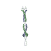
| Entry | Database: PDB / ID: 3s9g | ||||||
|---|---|---|---|---|---|---|---|
| Title | Structure of human Hexim1 (delta stammer) coiled coil domain | ||||||
 Components Components | Protein HEXIM1 | ||||||
 Keywords Keywords |  TRANSCRIPTION / Cyclin T-binding domain (TBD) / Cyclin T1/P-TEFb/7SK snRNA / nucleus TRANSCRIPTION / Cyclin T-binding domain (TBD) / Cyclin T1/P-TEFb/7SK snRNA / nucleus | ||||||
| Function / homology |  Function and homology information Function and homology information7SK snRNP / 7SK snRNA binding / snRNA binding / negative regulation of viral transcription / negative regulation of cyclin-dependent protein serine/threonine kinase activity / cyclin-dependent protein serine/threonine kinase inhibitor activity / protein kinase inhibitor activity / negative regulation of transcription elongation by RNA polymerase II / positive regulation of signal transduction by p53 class mediator / P-TEFb complex binding ...7SK snRNP / 7SK snRNA binding / snRNA binding / negative regulation of viral transcription / negative regulation of cyclin-dependent protein serine/threonine kinase activity / cyclin-dependent protein serine/threonine kinase inhibitor activity / protein kinase inhibitor activity / negative regulation of transcription elongation by RNA polymerase II / positive regulation of signal transduction by p53 class mediator / P-TEFb complex binding / activation of innate immune response / heart development / intracellular membrane-bounded organelle / innate immune response / negative regulation of DNA-templated transcription / negative regulation of transcription by RNA polymerase II / nucleoplasm / identical protein binding / nucleus / cytoplasmSimilarity search - Function | ||||||
| Biological species |  Homo sapiens (human) Homo sapiens (human) | ||||||
| Method | X-RAY DIFFRACTION / SYNCHROTRON / SAD / Resolution: 2.1 Å | ||||||
 Authors Authors | Bigalke, J.M. / Blankenfeldt, W. / Geyer, M. | ||||||
 Citation Citation | Journal: J.Mol.Biol. / Year: 2011 Title: Structure and Dynamics of a Stabilized Coiled-Coil Domain in the P-TEFb Regulator Hexim1. Authors: Bigalke, J.M. / Dames, S.A. / Blankenfeldt, W. / Grzesiek, S. / Geyer, M. | ||||||
| History |
|
- Structure visualization
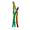
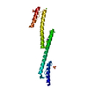
| Structure viewer | Molecule: MolmilJmol/JSmol |
|---|
- Downloads & links
-Download
| PDBx/mmCIF format | 3s9g.cif.gz | 78.6 KB | Display | PDBx/mmCIF format |
|---|---|---|---|---|
| PDB format | pdb3s9g.ent.gz | 60.5 KB | Display | PDB format |
| PDBx/mmJSON format | 3s9g.json.gz | Tree view | PDBx/mmJSON format | |
| Others |  Other downloads Other downloads |
-Validation report
| Arichive directory | https://data.pdbj.org/pub/pdb/validation_reports/s9/3s9gftp://data.pdbj.org/pub/pdb/validation_reports/s9/3s9g | HTTPS FTP |
|---|
-Related structure data
| Related structure data | |
|---|---|
| Similar structure data |


-Links
 PDBj
PDBj- Assembly
| Deposited unit | 
| ||||||||
|---|---|---|---|---|---|---|---|---|---|
| 1 |
| ||||||||
| Unit cell |
|
-Components
| #1: Protein | Mass: 12420.767 Da / Num. of mol.: 2 / Fragment: Cyclin T-binding domain / Mutation: C297S, delta(316-318) Source method: isolated from a genetically manipulated source Source: (gene. exp.) Homo sapiens (human) / Gene: CLP1, EDG1, HEXIM1, HIS1, MAQ1 / Plasmid: pProEx-HTa / Production host:  Escherichia coli (E. coli) / Strain (production host): BL21(DE3) / References: UniProt: O94992 Escherichia coli (E. coli) / Strain (production host): BL21(DE3) / References: UniProt: O94992#2: Water | ChemComp-HOH / | Water Mass: 18.015 Da / Num. of mol.: 51 / Source method: isolated from a natural source / Formula: H2O Mass: 18.015 Da / Num. of mol.: 51 / Source method: isolated from a natural source / Formula: H2O |
|---|
-Experimental details
-Experiment
| Experiment | Method: X-RAY DIFFRACTION / Number of used crystals: 1 |
|---|
- Sample preparation
| Crystal | Density Matthews: 2.29 Å3/Da / Density % sol: 46.34 % |
|---|---|
| Crystal grow | Temperature: 293 K / Method: vapor diffusion, hanging drop / pH: 5.5 Details: 0.1 M sodium cacodylate, 0.1 M calcium acetate, 4% PEG 8000, 5 mM magnesium chloride, pH 5.5, VAPOR DIFFUSION, HANGING DROP, temperature 293K |
-Data collection
| Diffraction | Mean temperature: 100 K |
|---|---|
| Diffraction source | Source: SYNCHROTRON / Site: SLS  / Beamline: X10SA / Wavelength: 0.97943 Å / Beamline: X10SA / Wavelength: 0.97943 Å |
| Detector | Type: MARMOSAIC 225 mm CCD / Detector: CCD / Date: Jun 30, 2007 |
| Radiation | Monochromator: Diamond(111) / Protocol: SINGLE WAVELENGTH / Monochromatic (M) / Laue (L): M / Scattering type: x-ray |
| Radiation wavelength | Wavelength: 0.97943 Å / Relative weight: 1 |
| Reflection | Resolution: 2.1→45.5 Å / Num. obs: 13117 / % possible obs: 100 % / Observed criterion σ(F): -3 / Observed criterion σ(I): -3 / Rmerge(I) obs: 0.085 / Net I/σ(I): 14.7 |
| Reflection shell | Resolution: 2.1→2.15 Å / Rmerge(I) obs: 0.386 / Mean I/σ(I) obs: 3.55 / % possible all: 100 |
- Processing
| Software |
| ||||||||||||||||||||||||||||||||||||||||||||||||||||||||||||||||||||||||||||||||||||||||||||||||||||||||||||||||||||||||||||||||||||||||||||||||||||||||||||||||||||||||||
|---|---|---|---|---|---|---|---|---|---|---|---|---|---|---|---|---|---|---|---|---|---|---|---|---|---|---|---|---|---|---|---|---|---|---|---|---|---|---|---|---|---|---|---|---|---|---|---|---|---|---|---|---|---|---|---|---|---|---|---|---|---|---|---|---|---|---|---|---|---|---|---|---|---|---|---|---|---|---|---|---|---|---|---|---|---|---|---|---|---|---|---|---|---|---|---|---|---|---|---|---|---|---|---|---|---|---|---|---|---|---|---|---|---|---|---|---|---|---|---|---|---|---|---|---|---|---|---|---|---|---|---|---|---|---|---|---|---|---|---|---|---|---|---|---|---|---|---|---|---|---|---|---|---|---|---|---|---|---|---|---|---|---|---|---|---|---|---|---|---|---|---|
| Refinement | Method to determine structure: SAD / Resolution: 2.1→45.5 Å / Cor.coef. Fo:Fc: 0.937 / Cor.coef. Fo:Fc free: 0.904 / SU B: 17.536 / SU ML: 0.203 / Cross valid method: THROUGHOUT / ESU R: 0.216 / ESU R Free: 0.205 / Stereochemistry target values: MAXIMUM LIKELIHOOD / Details: HYDROGENS HAVE BEEN ADDED IN THE RIDING POSITIONS
| ||||||||||||||||||||||||||||||||||||||||||||||||||||||||||||||||||||||||||||||||||||||||||||||||||||||||||||||||||||||||||||||||||||||||||||||||||||||||||||||||||||||||||
| Solvent computation | Ion probe radii: 0.8 Å / Shrinkage radii: 0.8 Å / VDW probe radii: 1.4 Å / Solvent model: BABINET MODEL WITH MASK | ||||||||||||||||||||||||||||||||||||||||||||||||||||||||||||||||||||||||||||||||||||||||||||||||||||||||||||||||||||||||||||||||||||||||||||||||||||||||||||||||||||||||||
| Displacement parameters | Biso mean: 52.557 Å2
| ||||||||||||||||||||||||||||||||||||||||||||||||||||||||||||||||||||||||||||||||||||||||||||||||||||||||||||||||||||||||||||||||||||||||||||||||||||||||||||||||||||||||||
| Refinement step | Cycle: LAST / Resolution: 2.1→45.5 Å
| ||||||||||||||||||||||||||||||||||||||||||||||||||||||||||||||||||||||||||||||||||||||||||||||||||||||||||||||||||||||||||||||||||||||||||||||||||||||||||||||||||||||||||
| Refine LS restraints |
| ||||||||||||||||||||||||||||||||||||||||||||||||||||||||||||||||||||||||||||||||||||||||||||||||||||||||||||||||||||||||||||||||||||||||||||||||||||||||||||||||||||||||||
| LS refinement shell | Resolution: 2.1→2.155 Å / Total num. of bins used: 20
| ||||||||||||||||||||||||||||||||||||||||||||||||||||||||||||||||||||||||||||||||||||||||||||||||||||||||||||||||||||||||||||||||||||||||||||||||||||||||||||||||||||||||||
| Refinement TLS params. | Method: refined / Refine-ID: X-RAY DIFFRACTION
| ||||||||||||||||||||||||||||||||||||||||||||||||||||||||||||||||||||||||||||||||||||||||||||||||||||||||||||||||||||||||||||||||||||||||||||||||||||||||||||||||||||||||||
| Refinement TLS group |
|
