Mass: 18.015 Da / Num. of mol.: 21 / Source method: isolated from a natural source / Formula: H2O
-
Experimental details
-
Experiment
Experiment
Method: X-RAY DIFFRACTION / Number of used crystals: 1
-
Sample preparation
Crystal
ID
Density Matthews (Å3/Da)
Density % sol (%)
1
2.59
52.54
2
3
Crystal grow
Temperature (K)
Crystal-ID
Details
PH range
291
1
crystal used for final refinement: 1.5 M ammonium phosphate, 0.1 M bis-tris propane. 1:200 trypsin was also added., pH 8.5, vapor diffusion, temperature 291K
8.5; 7; 6.5
291
2
Selenomethionyl derivative: 1.5 M ammonium phosphat e, 0.1 M bis-tris propane. 1:200 trypsin was also added., pH 7, vapor diffusion, temperature 291K
291
3
crystal used for preliminary refinement: 25 w/v% PEG-3350, 0.2 M lithium sulfate, 0.1 M bis-tris propane. 1:200 trypsin was also added., pH 6.5, vapor diffusion, temperature 291K
-
Data collection
Diffraction
ID
Mean temperature (K)
Crystal-ID
1
100
1
2
100
2
3
100
3
Diffraction source
Source
Site
Beamline
Type
ID
Wavelength
ROTATING ANODE
RIGAKU FR-E
1
1.5418
SYNCHROTRON
CHESS
A1
2
0.977
SYNCHROTRON
CLSI
08ID-1
3
0.97949
Detector
Type
ID
Detector
Date
RIGAKU SATURN
1
CCD
Apr 18, 2011
ADSC Q210 BINNED
2
CCD
May 11, 2011
MAR 300
3
CCD
May 25, 2011
Radiation
ID
Protocol
Monochromatic (M) / Laue (L)
Scattering type
Wavelength-ID
1
SINGLEWAVELENGTH
M
x-ray
1
2
SINGLEWAVELENGTH
M
x-ray
1
3
SINGLEWAVELENGTH
M
x-ray
1
Radiation wavelength
ID
Wavelength (Å)
Relative weight
1
1.5418
1
2
0.977
1
3
0.97949
1
Reflection
Resolution: 2.28→30 Å / Num. obs: 10833 / % possible obs: 94.8 % / Observed criterion σ(I): -3 / Biso Wilson estimate: 50.51 Å2 / Rmerge(I) obs: 0.038 / Net I/σ(I): 33.8
Reflection shell
Resolution: 2.28→2.34 Å / Rmerge(I) obs: 0.791 / Mean I/σ(I) obs: 2.8 / % possible all: 76.9
-
Phasing
Phasing
Method: SAD
-
Processing
Software
Name
Version
Classification
NB
XSCALE
datascaling
SOLVE
phasing
RESOLVE
phasing
BUSTER-TNT
BUSTER2.8.0
refinement
PDB_EXTRACT
3.1
dataextraction
BUSTER
2.8.0
refinement
Refinement
Method to determine structure: SAD / Resolution: 2.28→27.48 Å / Cor.coef. Fo:Fc: 0.91 / Cor.coef. Fo:Fc free: 0.887 / Occupancy max: 1 / Occupancy min: 0.5 / Cross valid method: THROUGHOUT / σ(F): 0 Details: BUCCANEER AND ARP/WARP WERE USED FOR AUTOMATED MODEL TRACING. DM WAS USED FOR ADDITIONAL PHASE REFINEMENT. REFMAC AND PHENIX WERE ALSO USED FOR REFINEMENT. COOT WAS USED FOR INTERACTIVE ...Details: BUCCANEER AND ARP/WARP WERE USED FOR AUTOMATED MODEL TRACING. DM WAS USED FOR ADDITIONAL PHASE REFINEMENT. REFMAC AND PHENIX WERE ALSO USED FOR REFINEMENT. COOT WAS USED FOR INTERACTIVE MODEL COMPLETION. THE MOLPROBITY SERVER WAS USED TO EVALUATE MODEL GEOMETRY. ABOUT THE DATA SETS: (NATIVE) DATA SET 1 WAS USED FOR FINAL REFINEMENT. OF THE AVAILABLE DATA SETS, SET ONE EXTENDED TO THE HIGHEST RESOLUTION. HOWEVER, DIFFRACTION IMAGES HAD ICE RINGS AND COMPLETENESS WAS < 95%. DATA SET 2 (P212121; A,B,C = 43.00A,43.56A,126.53A; PROCESSED WITH HKL2000) WAS USED FOR SELENIUM-SAD PHASING. (NATIVE) DATA SET 3 (P212121; A,B,C = 42.83,43.38,126.72A; PROCESSED WITH XDS) IS COMPLETE TO 2.5 A RESOLUTION AND WAS USED DURING INTERMEDIATE STAGES OF MODEL REFINEMENT.
Rfactor
Num. reflection
% reflection
Selection details
Rfree
0.271
575
5.32 %
THIN SHELLS (SFTOOLS)
Rwork
0.245
-
-
-
obs
0.246
10803
-
-
Displacement parameters
Biso mean: 48.56 Å2
Baniso -1
Baniso -2
Baniso -3
1-
2.5105 Å2
0 Å2
0 Å2
2-
-
-3.3032 Å2
0 Å2
3-
-
-
0.7928 Å2
Refine analyze
Luzzati coordinate error obs: 0.47 Å
Refinement step
Cycle: LAST / Resolution: 2.28→27.48 Å
Protein
Nucleic acid
Ligand
Solvent
Total
Num. atoms
1197
0
8
21
1226
Refine LS restraints
Refine-ID
Type
Dev ideal
Number
Restraint function
Weight
X-RAY DIFFRACTION
t_bond_d
0.01
1225
HARMONIC
2
X-RAY DIFFRACTION
t_angle_deg
1.06
1661
HARMONIC
2
X-RAY DIFFRACTION
t_dihedral_angle_d
444
SINUSOIDAL
2
X-RAY DIFFRACTION
t_incorr_chiral_ct
X-RAY DIFFRACTION
t_pseud_angle
X-RAY DIFFRACTION
t_trig_c_planes
23
HARMONIC
2
X-RAY DIFFRACTION
t_gen_planes
181
HARMONIC
5
X-RAY DIFFRACTION
t_it
1225
HARMONIC
20
X-RAY DIFFRACTION
t_nbd
X-RAY DIFFRACTION
t_omega_torsion
2.61
X-RAY DIFFRACTION
t_other_torsion
17.08
X-RAY DIFFRACTION
t_improper_torsion
X-RAY DIFFRACTION
t_chiral_improper_torsion
168
SEMIHARMONIC
5
X-RAY DIFFRACTION
t_sum_occupancies
X-RAY DIFFRACTION
t_utility_distance
X-RAY DIFFRACTION
t_utility_angle
X-RAY DIFFRACTION
t_utility_torsion
X-RAY DIFFRACTION
t_ideal_dist_contact
1422
SEMIHARMONIC
4
LS refinement shell
Resolution: 2.28→2.55 Å / Total num. of bins used: 5
Rfactor
Num. reflection
% reflection
Rfree
0.3058
124
4.24 %
Rwork
0.2824
2798
-
all
0.2835
2922
-
Refinement TLS params.
Method: refined / Refine-ID: X-RAY DIFFRACTION
ID
L11 (°2)
L12 (°2)
L13 (°2)
L22 (°2)
L23 (°2)
L33 (°2)
S11 (Å °)
S12 (Å °)
S13 (Å °)
S21 (Å °)
S22 (Å °)
S23 (Å °)
S31 (Å °)
S32 (Å °)
S33 (Å °)
T11 (Å2)
T12 (Å2)
T13 (Å2)
T22 (Å2)
T23 (Å2)
T33 (Å2)
Origin x (Å)
Origin y (Å)
Origin z (Å)
1
2.8595
1.1223
-2.383
3.5422
-0.245
7.0788
0.0408
-0.0665
0.2378
0.5479
0.1196
0.5254
-0.0057
-0.5269
-0.1604
0.0276
0.0002
0.0746
-0.1684
0.0363
-0.1627
14.8015
8.0628
29.6696
2
3.6242
0.1422
-2.3236
0.9209
-0.3711
4.9753
0.0085
0.2356
-0.3681
-0.2115
-0.0282
-0.015
0.6665
-0.119
0.0198
-0.0317
-0.0282
-0.0366
-0.0416
-0.0398
-0.1699
22.0078
3.6844
-0.3128
Refinement TLS group
ID
Refine-ID
Refine TLS-ID
Selection details
Auth asym-ID
Auth seq-ID
1
X-RAY DIFFRACTION
1
{ A|* }
A
0 - 79
2
X-RAY DIFFRACTION
2
{ B|* }
B
0 - 80
+
About Yorodumi
-
News
-
Feb 9, 2022. New format data for meta-information of EMDB entries
New format data for meta-information of EMDB entries
Version 3 of the EMDB header file is now the official format.
The previous official version 1.9 will be removed from the archive.
In the structure databanks used in Yorodumi, some data are registered as the other names, "COVID-19 virus" and "2019-nCoV". Here are the details of the virus and the list of structure data.
Jan 31, 2019. EMDB accession codes are about to change! (news from PDBe EMDB page)
EMDB accession codes are about to change! (news from PDBe EMDB page)
The allocation of 4 digits for EMDB accession codes will soon come to an end. Whilst these codes will remain in use, new EMDB accession codes will include an additional digit and will expand incrementally as the available range of codes is exhausted. The current 4-digit format prefixed with “EMD-” (i.e. EMD-XXXX) will advance to a 5-digit format (i.e. EMD-XXXXX), and so on. It is currently estimated that the 4-digit codes will be depleted around Spring 2019, at which point the 5-digit format will come into force.
The EM Navigator/Yorodumi systems omit the EMD- prefix.
Related info.:Q: What is EMD? / ID/Accession-code notation in Yorodumi/EM Navigator
Yorodumi is a browser for structure data from EMDB, PDB, SASBDB, etc.
This page is also the successor to EM Navigator detail page, and also detail information page/front-end page for Omokage search.
The word "yorodu" (or yorozu) is an old Japanese word meaning "ten thousand". "mi" (miru) is to see.
Related info.:EMDB / PDB / SASBDB / Comparison of 3 databanks / Yorodumi Search / Aug 31, 2016. New EM Navigator & Yorodumi / Yorodumi Papers / Jmol/JSmol / Function and homology information / Changes in new EM Navigator and Yorodumi
 Movie
Movie Controller


 Open data
 Basic information
 Components
 Keywords
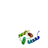
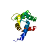
 TRANSCRIPTION /
  Function and homology information
 Authors
 Citation
 Structure visualization
 Downloads & links
 Other downloads


 PDBj
PDBj Assembly
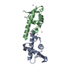


 Num. of mol.: 8 / Source method: obtained synthetically
 Mass: 18.015 Da / Num. of mol.: 21 / Source method: isolated from a natural source / Formula: H2O
 Sample preparation

 Processing