 Movie
Movie Controller


+ Open data

- Basic information
| Entry | Database: PDB / ID: 3s4u | ||||||
|---|---|---|---|---|---|---|---|
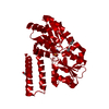
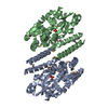
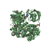
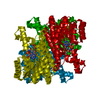
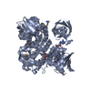
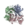
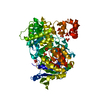
| Title | Crystal structure of open, unliganded E. coli PhnD H157A | ||||||
 Components Components | PhnD, subunit of alkylphosphonate ABC transporter | ||||||
 Keywords Keywords |  TRANSPORT PROTEIN / Phosphonate binding / Globular protein TRANSPORT PROTEIN / Phosphonate binding / Globular protein | ||||||
| Function / homology |  Function and homology information Function and homology informationorganic phosphonate transport / ATP-binding cassette (ABC) transporter complex / transmembrane transport Similarity search - Function | ||||||
| Biological species |  Escherichia coli (E. coli) Escherichia coli (E. coli) | ||||||
| Method | X-RAY DIFFRACTION / SYNCHROTRON / MOLECULAR REPLACEMENT / Resolution: 3.3 Å | ||||||
 Authors Authors | Alicea, I. / Schreiter, E.R. | ||||||
 Citation Citation | Journal: J.Mol.Biol. / Year: 2011 Title: Structure of the Escherichia coli Phosphonate Binding Protein PhnD and Rationally Optimized Phosphonate Biosensors. Authors: Alicea, I. / Marvin, J.S. / Miklos, A.E. / Ellington, A.D. / Looger, L.L. / Schreiter, E.R. | ||||||
| History |
|
- Structure visualization
| Structure viewer | Molecule: MolmilJmol/JSmol |
|---|
- Downloads & links
-Download
| PDBx/mmCIF format | 3s4u.cif.gz | 130 KB | Display | PDBx/mmCIF format |
|---|---|---|---|---|
| PDB format | pdb3s4u.ent.gz | 102.9 KB | Display | PDB format |
| PDBx/mmJSON format | 3s4u.json.gz | Tree view | PDBx/mmJSON format | |
| Others |  Other downloads Other downloads |
-Validation report
| Arichive directory | https://data.pdbj.org/pub/pdb/validation_reports/s4/3s4uftp://data.pdbj.org/pub/pdb/validation_reports/s4/3s4u | HTTPS FTP |
|---|
-Related structure data


-Links
 PDBj
PDBj- Assembly
| Deposited unit | 
| ||||||||
|---|---|---|---|---|---|---|---|---|---|
| 1 | 
| ||||||||
| 2 |
| ||||||||
| Unit cell |
|
-Components
| #1: Protein | Mass: 35741.379 Da / Num. of mol.: 1 / Fragment: UNP residues 27-338 / Mutation: H157A Source method: isolated from a genetically manipulated source Source: (gene. exp.) Escherichia coli (E. coli) / Strain: UTI89 / UPEC / Gene: phnD, UTI89_C4699 / Production host: Escherichia coli (E. coli) / Strain (production host): Bl21(DE3) / References: UniProt: Q1R3F7 |
|---|
-Experimental details
-Experiment
| Experiment | Method: X-RAY DIFFRACTION / Number of used crystals: 1 |
|---|
- Sample preparation
| Crystal | Density Matthews: 2.2 Å3/Da / Density % sol: 44.06 % |
|---|---|
| Crystal grow | Temperature: 298 K / Method: vapor diffusion, sitting drop / pH: 8.5 Details: 0.2 M magnesium chloride, 0.1 M Tris, pH 8.5, 25% w/v PEG 3350, VAPOR DIFFUSION, SITTING DROP, temperature 298K |
-Data collection
| Diffraction | Mean temperature: 298 K |
|---|---|
| Diffraction source | Source: SYNCHROTRON / Site: APS  / Beamline: 31-ID / Wavelength: 0.97929 Å / Beamline: 31-ID / Wavelength: 0.97929 Å |
| Detector | Type: RAYONIX MX225HE / Detector: CCD / Date: Aug 20, 2010 |
| Radiation | Monochromator: Diamond 111 / Protocol: SINGLE WAVELENGTH / Monochromatic (M) / Laue (L): M / Scattering type: x-ray |
| Radiation wavelength | Wavelength: 0.97929 Å / Relative weight: 1 |
| Reflection | Resolution: 3.3→20 Å / Num. all: 5149 / Num. obs: 5123 / % possible obs: 99.5 % / Observed criterion σ(F): 0 / Observed criterion σ(I): 0 |
- Processing
| Software |
| ||||||||||||||||||||||||||||||||||||||||||||||||||||||||||||
|---|---|---|---|---|---|---|---|---|---|---|---|---|---|---|---|---|---|---|---|---|---|---|---|---|---|---|---|---|---|---|---|---|---|---|---|---|---|---|---|---|---|---|---|---|---|---|---|---|---|---|---|---|---|---|---|---|---|---|---|---|---|
| Refinement | Method to determine structure: MOLECULAR REPLACEMENT / Resolution: 3.3→20 Å / Cor.coef. Fo:Fc: 0.926 / Cor.coef. Fo:Fc free: 0.824 / SU B: 70.628 / SU ML: 0.532 / Cross valid method: THROUGHOUT / σ(F): 0 / ESU R Free: 0.676 / Stereochemistry target values: MAXIMUM LIKELIHOOD / Details: HYDROGENS HAVE BEEN USED IF PRESENT IN THE INPUT
| ||||||||||||||||||||||||||||||||||||||||||||||||||||||||||||
| Solvent computation | Ion probe radii: 0.8 Å / Shrinkage radii: 0.8 Å / VDW probe radii: 1.2 Å / Solvent model: MASK | ||||||||||||||||||||||||||||||||||||||||||||||||||||||||||||
| Displacement parameters | Biso mean: 53 Å2
| ||||||||||||||||||||||||||||||||||||||||||||||||||||||||||||
| Refinement step | Cycle: LAST / Resolution: 3.3→20 Å
| ||||||||||||||||||||||||||||||||||||||||||||||||||||||||||||
| Refine LS restraints |
| ||||||||||||||||||||||||||||||||||||||||||||||||||||||||||||
| LS refinement shell | Resolution: 3.3→3.383 Å / Total num. of bins used: 20
| ||||||||||||||||||||||||||||||||||||||||||||||||||||||||||||
| Refinement TLS params. | Method: refined / Origin x: -5.836 Å / Origin y: -27.162 Å / Origin z: -5.571 Å
|
