 Movie
Movie Controller


+ Open data

- Basic information
| Entry | Database: PDB / ID: 3s3s | ||||||
|---|---|---|---|---|---|---|---|
| Title | Transglutaminase 2 in complex with a novel inhibitor | ||||||
 Components Components |
| ||||||
 Keywords Keywords | TRANSFERASE/TRANSFERASE INHIBITOR /  Transglutaminase / TRANSFERASE-TRANSFERASE INHIBITOR complex Transglutaminase / TRANSFERASE-TRANSFERASE INHIBITOR complex | ||||||
| Function / homology |  Function and homology information Function and homology informationhistone serotonyltransferase activity / histone dopaminyltransferase activity / peptide noradrenalinyltransferase activity / peptide histaminyltransferase activity / cellular response to serotonin / regulation of apoptotic cell clearance / protein deamination / protein-glutamine glutaminase activity / protein-glutamine glutaminase / protein-glutamine gamma-glutamyltransferase ...histone serotonyltransferase activity / histone dopaminyltransferase activity / peptide noradrenalinyltransferase activity / peptide histaminyltransferase activity / cellular response to serotonin / regulation of apoptotic cell clearance / protein deamination / protein-glutamine glutaminase activity / protein-glutamine glutaminase / protein-glutamine gamma-glutamyltransferase / positive regulation of mitochondrial calcium ion concentration / salivary gland cavitation / protein-glutamine gamma-glutamyltransferase activity / negative regulation of endoplasmic reticulum calcium ion concentration / dopamine secretion / peptide cross-linking / branching involved in salivary gland morphogenesis / cellular response to dopamine / positive regulation of small GTPase mediated signal transduction / Hydrolases; Acting on peptide bonds (peptidases) / apoptotic cell clearance / cellular response to cocaine / positive regulation of neurogenesis / positive regulation of cell adhesion / Transferases; Acyltransferases; Transferring groups other than aminoacyl groups / extracellular matrix / bone development / protein homooligomerization / positive regulation of GTPase activity / nucleosome / phospholipase C-activating G protein-coupled receptor signaling pathway / gene expression / peptidase activity / collagen-containing extracellular matrix / regulation of apoptotic process / positive regulation of apoptotic process / focal adhesion / calcium ion binding / chromatin / GTP binding / perinuclear region of cytoplasm / endoplasmic reticulum / mitochondrion / proteolysis / extracellular exosome / nucleus / plasma membrane / cytosolSimilarity search - Function | ||||||
| Biological species |  Homo sapiens (human) Homo sapiens (human)synthetic construct (others) | ||||||
| Method | X-RAY DIFFRACTION / SYNCHROTRON / MOLECULAR REPLACEMENT / Resolution: 2.301 Å | ||||||
 Authors Authors | Lindemann, I. / Heine, A. / Klebe, G. | ||||||
 Citation Citation | Journal: To be Published Title: Inhibitors of Transglutaminase 2: A therapeutic option in celiac disease Authors: Lindemann, I. / Boettcher, J. / Oertel, K. / Weber, J. / Hils, M. / Pasternack, R. / Heine, A. / Klebe, G. | ||||||
| History |
|
- Structure visualization
| Structure viewer | Molecule: MolmilJmol/JSmol |
|---|
- Downloads & links
-Download
| PDBx/mmCIF format | 3s3s.cif.gz | 141.2 KB | Display | PDBx/mmCIF format |
|---|---|---|---|---|
| PDB format | pdb3s3s.ent.gz | 107.2 KB | Display | PDB format |
| PDBx/mmJSON format | 3s3s.json.gz | Tree view | PDBx/mmJSON format | |
| Others |  Other downloads Other downloads |
-Validation report
| Arichive directory | https://data.pdbj.org/pub/pdb/validation_reports/s3/3s3sftp://data.pdbj.org/pub/pdb/validation_reports/s3/3s3s | HTTPS FTP |
|---|
-Related structure data
| Related structure data |  3s3jC  2q3zS S: Starting model for refinement C: citing same article ( |
|---|---|
| Similar structure data |


-Links
 PDBj
PDBj
- Assembly
| Deposited unit | 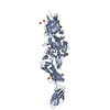
| ||||||||
|---|---|---|---|---|---|---|---|---|---|
| 1 |
| ||||||||
| Unit cell |
| ||||||||
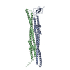
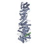
| Details | THE MAIN POLYMERIC PROTEIN-GLUTAMINE GAMMA-GLUTAMYLTRANSFERASE 2 IS A MONOMER IN THE ABSENCE OF THE PEPTIDE INHIBITOR |
-Components
| #1: Protein | Transglutaminase / Tissue transglutaminase / Transglutaminase C / TG(C) / TGC / TGase C / Transglutaminase H / TGase H ...Tissue transglutaminase / Transglutaminase C / TG(C) / TGC / TGase C / Transglutaminase H / TGase H / Transglutaminase-2 / TGase-2 Mass: 78312.641 Da / Num. of mol.: 1 / Fragment: unp residues 2-687 Source method: isolated from a genetically manipulated source Source: (gene. exp.) Homo sapiens (human) / Gene: TGM2 / Production host:  Escherichia coli (E. coli) Escherichia coli (E. coli)References: UniProt: P21980, protein-glutamine gamma-glutamyltransferase | ||||
|---|---|---|---|---|---|
| #2: Protein/peptide |   Type: Peptide-like / Class: Inhibitor / Mass: 665.217 Da / Num. of mol.: 1 / Source method: obtained synthetically / Source: (synth.) synthetic construct (others) Type: Peptide-like / Class: Inhibitor / Mass: 665.217 Da / Num. of mol.: 1 / Source method: obtained synthetically / Source: (synth.) synthetic construct (others)References: N-[(2R)-2-{[(benzyloxy)carbonyl]amino}-7-ethoxy-7-oxoheptanoyl]-L-valyl-L-prolyl-L-leucine | ||||
| #3: Chemical | ChemComp-SO4 / Sulfate  Mass: 96.063 Da / Num. of mol.: 6 / Source method: obtained synthetically / Formula: SO4 Mass: 96.063 Da / Num. of mol.: 6 / Source method: obtained synthetically / Formula: SO4#4: Water | ChemComp-HOH / | Water Mass: 18.015 Da / Num. of mol.: 153 / Source method: isolated from a natural source / Formula: H2O Mass: 18.015 Da / Num. of mol.: 153 / Source method: isolated from a natural source / Formula: H2OCompound details | THE UNREACTED FORM OF THE PEPTIDE INHIBITOR, CHAIN B HAS A DOUBLE BOND BETWEEN C15 AND C17 OF THE ...THE UNREACTED FORM OF THE PEPTIDE INHIBITOR, CHAIN B HAS A DOUBLE BOND BETWEEN C15 AND C17 OF THE AMINOACID XW1. UPON REACTION WITH PROTEIN, A COVALENT BOND BETWEEN C15 AND SG OF CYS 277 CHAIN A IS FORMED | |
-Experimental details
-Experiment
| Experiment | Method: X-RAY DIFFRACTION / Number of used crystals: 1 |
|---|
- Sample preparation
| Crystal | Density Matthews: 2.51 Å3/Da / Density % sol: 50.93 % |
|---|---|
| Crystal grow | Temperature: 289 K / Method: vapor diffusion, sitting drop / pH: 7 Details: 3.0 M Amm. sulfate, 0.1 M HEPES, VAPOR DIFFUSION, SITTING DROP, temperature 289K, pH 7.0 |
-Data collection
| Diffraction | Mean temperature: 100 K |
|---|---|
| Diffraction source | Source: SYNCHROTRON / Site: BESSY  / Beamline: 14.2 / Wavelength: 0.91841 Å / Beamline: 14.2 / Wavelength: 0.91841 Å |
| Detector | Type: RAYONIX MX-225 / Detector: CCD / Date: Oct 21, 2010 / Details: mirrors |
| Radiation | Monochromator: Double Crystal Monochromator KMC-2 / Protocol: SINGLE WAVELENGTH / Monochromatic (M) / Laue (L): M / Scattering type: x-ray |
| Radiation wavelength | Wavelength: 0.91841 Å / Relative weight: 1 |
| Reflection | Resolution: 2.3→50 Å / Num. all: 34641 / Num. obs: 34641 / % possible obs: 100 % / Redundancy: 7 % / Rsym value: 0.075 / Net I/σ(I): 23.5 |
| Reflection shell | Resolution: 2.3→2.35 Å / Redundancy: 4.8 % / Mean I/σ(I) obs: 3.5 / Num. unique all: 2006 / Rsym value: 0.345 / % possible all: 100 |
- Processing
| Software |
| |||||||||||||||||||||||||||||||||||||||||||||||||||||||||||||||||||||||||||||||||||||||||||
|---|---|---|---|---|---|---|---|---|---|---|---|---|---|---|---|---|---|---|---|---|---|---|---|---|---|---|---|---|---|---|---|---|---|---|---|---|---|---|---|---|---|---|---|---|---|---|---|---|---|---|---|---|---|---|---|---|---|---|---|---|---|---|---|---|---|---|---|---|---|---|---|---|---|---|---|---|---|---|---|---|---|---|---|---|---|---|---|---|---|---|---|---|
| Refinement | Method to determine structure: MOLECULAR REPLACEMENT Starting model: 2q3z Resolution: 2.301→48.034 Å / SU ML: 0.32 / σ(F): 0 / Phase error: 28.95 / Stereochemistry target values: ML
| |||||||||||||||||||||||||||||||||||||||||||||||||||||||||||||||||||||||||||||||||||||||||||
| Solvent computation | Shrinkage radii: 0.95 Å / VDW probe radii: 1.2 Å / Solvent model: FLAT BULK SOLVENT MODEL / Bsol: 62.497 Å2 / ksol: 0.359 e/Å3 | |||||||||||||||||||||||||||||||||||||||||||||||||||||||||||||||||||||||||||||||||||||||||||
| Displacement parameters |
| |||||||||||||||||||||||||||||||||||||||||||||||||||||||||||||||||||||||||||||||||||||||||||
| Refinement step | Cycle: LAST / Resolution: 2.301→48.034 Å
| |||||||||||||||||||||||||||||||||||||||||||||||||||||||||||||||||||||||||||||||||||||||||||
| Refine LS restraints |
| |||||||||||||||||||||||||||||||||||||||||||||||||||||||||||||||||||||||||||||||||||||||||||
| LS refinement shell |
|
