 Movie
Movie Controller


+ Open data

- Basic information
| Entry | Database: PDB / ID: 3ryl | ||||||
|---|---|---|---|---|---|---|---|
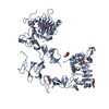
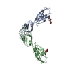
| Title | Dimerization domain of Vibrio parahemolyticus VopL | ||||||
 Components Components | protein VPA1370 | ||||||
 Keywords Keywords |  PROTEIN BINDING / Actin nucleation / Filament pointed end binding / Type III Secretion System (T3SS) protein PROTEIN BINDING / Actin nucleation / Filament pointed end binding / Type III Secretion System (T3SS) protein | ||||||
| Function / homology |  Function and homology information Function and homology information | ||||||
| Biological species |   Vibrio parahaemolyticus (bacteria) Vibrio parahaemolyticus (bacteria) | ||||||
| Method | X-RAY DIFFRACTION / SYNCHROTRON / SAD / Resolution: 3.1 Å | ||||||
 Authors Authors | Namgoong, S. / Dominguez, R. | ||||||
 Citation Citation | Journal: Nat.Struct.Mol.Biol. / Year: 2011 Title: Mechanism of actin filament nucleation by Vibrio VopL and implications for tandem W domain nucleation. Authors: Namgoong, S. / Boczkowska, M. / Glista, M.J. / Winkelman, J.D. / Rebowski, G. / Kovar, D.R. / Dominguez, R. | ||||||
| History |
|
- Structure visualization
| Structure viewer | Molecule: MolmilJmol/JSmol |
|---|
- Downloads & links
-Download
| PDBx/mmCIF format | 3ryl.cif.gz | 185.8 KB | Display | PDBx/mmCIF format |
|---|---|---|---|---|
| PDB format | pdb3ryl.ent.gz | 157.5 KB | Display | PDB format |
| PDBx/mmJSON format | 3ryl.json.gz | Tree view | PDBx/mmJSON format | |
| Others |  Other downloads Other downloads |
-Validation report
| Arichive directory | https://data.pdbj.org/pub/pdb/validation_reports/ry/3rylftp://data.pdbj.org/pub/pdb/validation_reports/ry/3ryl | HTTPS FTP |
|---|
-Related structure data
| Similar structure data |
|---|


-Links
 PDBj
PDBj- Assembly
| Deposited unit | 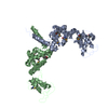
| ||||||||
|---|---|---|---|---|---|---|---|---|---|
| 1 |
| ||||||||
| Unit cell |
|
-Components
| #1: Protein | Mass: 27064.623 Da / Num. of mol.: 2 / Fragment: VopL dimerization domain (UNP residues 247-484) Source method: isolated from a genetically manipulated source Source: (gene. exp.) Vibrio parahaemolyticus (bacteria) / Strain: RIMD 2210633 / Gene: VopL, VPA1370 / Plasmid: pTYB12 / Production host: Escherichia coli (E. coli) / Strain (production host): BL21(DE3) / References: UniProt: Q87GE5 |
|---|
-Experimental details
-Experiment
| Experiment | Method: X-RAY DIFFRACTION / Number of used crystals: 1 |
|---|
- Sample preparation
| Crystal | Density Matthews: 2.42 Å3/Da / Density % sol: 49.25 % |
|---|---|
| Crystal grow | Temperature: 277 K / Method: vapor diffusion, hanging drop / pH: 7.5 Details: 13-15% PEG3350, 0.2 M sodium fluoride, pH 7.5, VAPOR DIFFUSION, HANGING DROP, temperature 277K |
-Data collection
| Diffraction | Mean temperature: 100 K |
|---|---|
| Diffraction source | Source: SYNCHROTRON / Site: CHESS  / Beamline: A1 / Wavelength: 0.9772 / Beamline: A1 / Wavelength: 0.9772 |
| Detector | Type: ADSC QUANTUM 210 / Detector: CCD / Date: Mar 29, 2011 / Details: Bent, triangular Si(111) monochromator crystal |
| Radiation | Monochromator: Si 111 CHANNEL / Protocol: SINGLE WAVELENGTH / Monochromatic (M) / Laue (L): M / Scattering type: x-ray |
| Radiation wavelength | Wavelength: 0.9772 Å / Relative weight: 1 |
| Reflection | Resolution: 3.1→67.65 Å / Num. all: 10025 / Num. obs: 10025 / % possible obs: 99.5 % / Observed criterion σ(F): 0 / Observed criterion σ(I): 0 / Redundancy: 32.75 % / Biso Wilson estimate: 107.97 Å2 / Rmerge(I) obs: 0.1224 / Rsym value: 0.1224 / Net I/σ(I): 20.56 |
| Reflection shell | Resolution: 3.1→3.2 Å / Redundancy: 34.86 % / Rmerge(I) obs: 0.4257 / Mean I/σ(I) obs: 2.37 / Num. unique all: 897 / Rsym value: 0.4257 / % possible all: 99.9 |
- Processing
| Software |
| ||||||||||||||||||||||||||||||||||||||||||||||||||||||||
|---|---|---|---|---|---|---|---|---|---|---|---|---|---|---|---|---|---|---|---|---|---|---|---|---|---|---|---|---|---|---|---|---|---|---|---|---|---|---|---|---|---|---|---|---|---|---|---|---|---|---|---|---|---|---|---|---|---|
| Refinement | Method to determine structure: SAD / Resolution: 3.1→67.65 Å / SU ML: 0.57 / σ(F): 0 / σ(I): 0 / Phase error: 29.51 / Stereochemistry target values: ML
| ||||||||||||||||||||||||||||||||||||||||||||||||||||||||
| Solvent computation | Shrinkage radii: 1.06 Å / VDW probe radii: 1.3 Å / Solvent model: FLAT BULK SOLVENT MODEL / Bsol: 118.768 Å2 / ksol: 0.3 e/Å3 | ||||||||||||||||||||||||||||||||||||||||||||||||||||||||
| Displacement parameters | Biso mean: 149.12 Å2
| ||||||||||||||||||||||||||||||||||||||||||||||||||||||||
| Refinement step | Cycle: LAST / Resolution: 3.1→67.65 Å
| ||||||||||||||||||||||||||||||||||||||||||||||||||||||||
| Refine LS restraints |
| ||||||||||||||||||||||||||||||||||||||||||||||||||||||||
| LS refinement shell | Refine-ID: X-RAY DIFFRACTION
|
