- PDB-3rwr: Crystal structure of the human XRCC4-XLF complex -
+
Open data
ID or keywords:
Loading...
-
Basic information
Entry
Database: PDB / ID: 3rwr
Title
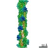
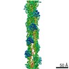
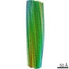
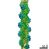
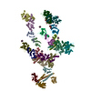
Crystal structure of the human XRCC4-XLF complex
Components
DNA repair protein XRCC4
Non-homologous end-joining factor 1
Keywords
DNA binding protein/protein binding / complex-filament / non-homologous end-joining / DNA and Protein Binding / DNA binding protein-protein binding complex
Function / homology
Function and homology information
FHA domain binding / positive regulation of ligase activity / DNA ligase IV complex / DNA ligation involved in DNA repair / DNA end binding / DNA-dependent protein kinase-DNA ligase 4 complex / immunoglobulin V(D)J recombination / nonhomologous end joining complex / protein localization to site of double-strand break / response to ionizing radiation ...FHA domain binding / positive regulation of ligase activity / DNA ligase IV complex / DNA ligation involved in DNA repair / DNA end binding / DNA-dependent protein kinase-DNA ligase 4 complex / immunoglobulin V(D)J recombination / nonhomologous end joining complex / protein localization to site of double-strand break / response to ionizing radiation / cellular response to lithium ion / 2-LTR circle formation / T cell differentiation / response to X-ray / SUMOylation of DNA damage response and repair proteins / DNA polymerase binding / B cell differentiation / central nervous system development / Nonhomologous End-Joining (NHEJ) / fibrillar center / double-strand break repair via nonhomologous end joining / double-strand break repair / site of double-strand break / nucleoplasm / identical protein binding / nucleus / cytosol Similarity search - Function
Helix hairpin bin / Single alpha-helices involved in coiled-coils or other helix-helix interfaces - #370 / DNA double-strand break repair and VJ recombination XRCC4, N-terminal / Dna Repair Protein Xrcc4; Chain: A, domain 1 / XLF, N-terminal / XLF N-terminal domain / XRCC4, N-terminal domain superfamily / DNA repair protein XRCC4 / XRCC4 N-terminal domain / XRCC4-like, N-terminal domain superfamily ...Helix hairpin bin / Single alpha-helices involved in coiled-coils or other helix-helix interfaces - #370 / DNA double-strand break repair and VJ recombination XRCC4, N-terminal / Dna Repair Protein Xrcc4; Chain: A, domain 1 / XLF, N-terminal / XLF N-terminal domain / XRCC4, N-terminal domain superfamily / DNA repair protein XRCC4 / XRCC4 N-terminal domain / XRCC4-like, N-terminal domain superfamily / DNA repair protein XRCC4-like, C-terminal / Beta Complex / Single alpha-helices involved in coiled-coils or other helix-helix interfaces / Helix Hairpins / Up-down Bundle / Orthogonal Bundle / Mainly Beta / Mainly Alpha Similarity search - Domain/homology
HEXATANTALUM DODECABROMIDE / DNA repair protein XRCC4 / Non-homologous end-joining factor 1 Similarity search - Component
The biological assembly is a filament of alternating homodimers of XRCC4 and XLF. The unit is assembled by the following chains: GF, IH, BA, ED. This has been confirmed by SAXS (Hammel, M., Yu, Y., Fang, S., Lees-Miller, S.P., and Tainer, J.A. (2010). XLF regulates filament architecture of the XRCC4-ligaseIV complex. Structure. 11, 1431-1442.).
-
Components
#1: Protein
DNArepairproteinXRCC4 / / X-ray repair cross-complementing protein 4
Mass: 18869.123 Da / Num. of mol.: 12 / Fragment: unp residues 1-157 Source method: isolated from a genetically manipulated source Source: (gene. exp.) Homo sapiens (human) / Gene: XRCC4 / Production host: Escherichia coli (E. coli) / Strain (production host): BL21 (DE3) / References: UniProt: Q13426
#2: Protein
Non-homologousend-joiningfactor1 / Protein cernunnos / XRCC4-like factor
Mass: 26286.348 Da / Num. of mol.: 12 / Fragment: unp residues 1-224 Source method: isolated from a genetically manipulated source Source: (gene. exp.) Homo sapiens (human) / Gene: NHEJ1, XLF / Production host: Escherichia coli (E. coli) / Strain (production host): Rosetta (DE3) / References: UniProt: Q9H9Q4
Method to determine structure: molecular replacement-SAD / Resolution: 3.943→49.027 Å / SU ML: 1.55 / σ(F): 0 / Phase error: 36.08 / Stereochemistry target values: MLHL Details: THE TA ATOM FOR CHAINS P AND V HAS BEEN REFINED WITH OCCUPANCY HIGHER THAN 1.0.
Rfactor
Num. reflection
% reflection
Rfree
0.3256
3879
5.03 %
Rwork
0.2707
-
-
obs
0.2735
77191
99.14 %
all
-
77252
-
Solvent computation
Shrinkage radii: 0.86 Å / VDW probe radii: 1.1 Å / Solvent model: FLAT BULK SOLVENT MODEL / Bsol: 157.435 Å2 / ksol: 0.318 e/Å3
Displacement parameters
Baniso -1
Baniso -2
Baniso -3
1-
-58.2565 Å2
0 Å2
-37.7716 Å2
2-
-
44.8642 Å2
-0 Å2
3-
-
-
13.3923 Å2
Refinement step
Cycle: LAST / Resolution: 3.943→49.027 Å
Protein
Nucleic acid
Ligand
Solvent
Total
Num. atoms
36689
0
144
0
36833
Refine LS restraints
Refine-ID
Type
Dev ideal
Number
X-RAY DIFFRACTION
f_bond_d
0.003
37747
X-RAY DIFFRACTION
f_angle_d
0.788
50670
X-RAY DIFFRACTION
f_dihedral_angle_d
19.53
13882
X-RAY DIFFRACTION
f_chiral_restr
0.065
5660
X-RAY DIFFRACTION
f_plane_restr
0.003
6519
LS refinement shell
Resolution (Å)
Rfactor Rfree
Num. reflection Rfree
Rfactor Rwork
Num. reflection Rwork
Refine-ID
% reflection obs (%)
3.9427-3.9907
0.4165
95
0.3931
2157
X-RAY DIFFRACTION
81
3.9907-4.0412
0.4149
151
0.3809
2644
X-RAY DIFFRACTION
100
4.0412-4.0944
0.4066
139
0.3621
2532
X-RAY DIFFRACTION
100
4.0944-4.1504
0.4285
137
0.3556
2683
X-RAY DIFFRACTION
100
4.1504-4.2097
0.3781
123
0.3287
2624
X-RAY DIFFRACTION
100
4.2097-4.2725
0.3545
152
0.3223
2631
X-RAY DIFFRACTION
100
4.2725-4.3392
0.3555
134
0.2915
2557
X-RAY DIFFRACTION
100
4.3392-4.4103
0.3134
128
0.2671
2660
X-RAY DIFFRACTION
100
4.4103-4.4863
0.3318
136
0.2591
2696
X-RAY DIFFRACTION
100
4.4863-4.5678
0.3209
120
0.2602
2602
X-RAY DIFFRACTION
100
4.5678-4.6556
0.2949
126
0.2376
2615
X-RAY DIFFRACTION
100
4.6556-4.7506
0.3181
149
0.2449
2665
X-RAY DIFFRACTION
100
4.7506-4.8538
0.3134
128
0.2379
2590
X-RAY DIFFRACTION
100
4.8538-4.9666
0.2896
134
0.2442
2647
X-RAY DIFFRACTION
100
4.9666-5.0907
0.3269
144
0.2498
2613
X-RAY DIFFRACTION
100
5.0907-5.2281
0.3129
123
0.252
2642
X-RAY DIFFRACTION
100
5.2281-5.3818
0.3989
150
0.2787
2681
X-RAY DIFFRACTION
100
5.3818-5.5553
0.3948
144
0.3402
2573
X-RAY DIFFRACTION
100
5.5553-5.7535
0.4465
164
0.367
2665
X-RAY DIFFRACTION
100
5.7535-5.9835
0.3853
140
0.3558
2584
X-RAY DIFFRACTION
100
5.9835-6.2553
0.4025
148
0.3676
2677
X-RAY DIFFRACTION
100
6.2553-6.5844
0.4378
146
0.3594
2611
X-RAY DIFFRACTION
100
6.5844-6.9958
0.3842
126
0.3276
2640
X-RAY DIFFRACTION
100
6.9958-7.5342
0.4006
142
0.2813
2667
X-RAY DIFFRACTION
100
7.5342-8.2891
0.2735
170
0.2102
2630
X-RAY DIFFRACTION
100
8.2891-9.4811
0.2229
145
0.1666
2665
X-RAY DIFFRACTION
100
9.4811-11.9167
0.2029
150
0.1714
2637
X-RAY DIFFRACTION
99
11.9167-49.0304
0.337
135
0.3011
2724
X-RAY DIFFRACTION
99
+
About Yorodumi
-
News
-
Feb 9, 2022. New format data for meta-information of EMDB entries
New format data for meta-information of EMDB entries
Version 3 of the EMDB header file is now the official format.
The previous official version 1.9 will be removed from the archive.
In the structure databanks used in Yorodumi, some data are registered as the other names, "COVID-19 virus" and "2019-nCoV". Here are the details of the virus and the list of structure data.
Jan 31, 2019. EMDB accession codes are about to change! (news from PDBe EMDB page)
EMDB accession codes are about to change! (news from PDBe EMDB page)
The allocation of 4 digits for EMDB accession codes will soon come to an end. Whilst these codes will remain in use, new EMDB accession codes will include an additional digit and will expand incrementally as the available range of codes is exhausted. The current 4-digit format prefixed with “EMD-” (i.e. EMD-XXXX) will advance to a 5-digit format (i.e. EMD-XXXXX), and so on. It is currently estimated that the 4-digit codes will be depleted around Spring 2019, at which point the 5-digit format will come into force.
The EM Navigator/Yorodumi systems omit the EMD- prefix.
Related info.:Q: What is EMD? / ID/Accession-code notation in Yorodumi/EM Navigator
Yorodumi is a browser for structure data from EMDB, PDB, SASBDB, etc.
This page is also the successor to EM Navigator detail page, and also detail information page/front-end page for Omokage search.
The word "yorodu" (or yorozu) is an old Japanese word meaning "ten thousand". "mi" (miru) is to see.
Related info.:EMDB / PDB / SASBDB / Comparison of 3 databanks / Yorodumi Search / Aug 31, 2016. New EM Navigator & Yorodumi / Yorodumi Papers / Jmol/JSmol / Function and homology information / Changes in new EM Navigator and Yorodumi
 Movie
Movie Controller


 Open data
 Basic information
 Components
 Keywords
 non-homologous end-joining / DNA and Protein Binding / DNA binding protein-protein binding complex
 Function and homology information
 Authors
 Citation
 Structure visualization
 Downloads & links
 Other downloads
 PDBj
PDBj


 Assembly

 Mass: 2044.535 Da / Num. of mol.: 8 / Source method: obtained synthetically / Formula: Br12Ta6
 Sample preparation
 / Beamline: X25 / Wavelength: 1.2536 Å
 Processing