 Movie
Movie Controller


+ Open data

- Basic information
| Entry | Database: PDB / ID: 3rwe | ||||||
|---|---|---|---|---|---|---|---|
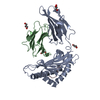
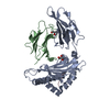
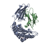
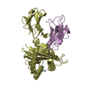
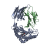
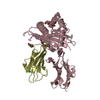
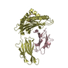
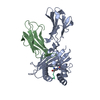
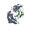
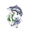
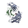
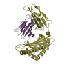
| Title | rhesus macaque MHC class I molecule Mamu-B*17-FW9 | ||||||
 Components Components |
| ||||||
 Keywords Keywords |  IMMUNE SYSTEM / antigenic peptides / T lymphocytes / immune response IMMUNE SYSTEM / antigenic peptides / T lymphocytes / immune response | ||||||
| Function / homology |  Function and homology information Function and homology informationantigen processing and presentation of peptide antigen via MHC class I / RNA stem-loop binding / antigen processing and presentation of endogenous peptide antigen via MHC class I via ER pathway, TAP-independent / antigen processing and presentation of endogenous peptide antigen via MHC class Ib / lumenal side of endoplasmic reticulum membrane / ER to Golgi transport vesicle membrane / exoribonuclease H activity / MHC class I protein complex / DNA integration / peptide antigen assembly with MHC class II protein complex ...antigen processing and presentation of peptide antigen via MHC class I / RNA stem-loop binding / antigen processing and presentation of endogenous peptide antigen via MHC class I via ER pathway, TAP-independent / antigen processing and presentation of endogenous peptide antigen via MHC class Ib / lumenal side of endoplasmic reticulum membrane / ER to Golgi transport vesicle membrane / exoribonuclease H activity / MHC class I protein complex / DNA integration / peptide antigen assembly with MHC class II protein complex / MHC class II protein complex / positive regulation of T cell mediated cytotoxicity / viral genome integration into host DNA / recycling endosome membrane / establishment of integrated proviral latency / phagocytic vesicle membrane / peptide antigen binding / antigen processing and presentation of exogenous peptide antigen via MHC class II / RNA-directed DNA polymerase activity / positive regulation of immune response / RNA-DNA hybrid ribonuclease activity / positive regulation of T cell activation / MHC class II protein complex binding / late endosome membrane / early endosome membrane / DNA recombination / aspartic-type endopeptidase activity / symbiont entry into host cell / immune response / lysosomal membrane / external side of plasma membrane / signaling receptor binding / proteolysis / DNA binding / extracellular space / zinc ion binding / extracellular regionSimilarity search - Function | ||||||
| Biological species |  Macaca mulatta (Rhesus monkey) Macaca mulatta (Rhesus monkey) Simian immunodeficiency virus Simian immunodeficiency virus | ||||||
| Method | X-RAY DIFFRACTION / MOLECULAR REPLACEMENT / Resolution: 2.4 Å | ||||||
 Authors Authors | Wu, Y. / Gao, F. / Liu, J. / Qi, J.X. / Price, D.A. / Gao, G.F. | ||||||
 Citation Citation | Journal: J.Immunol. / Year: 2011 Title: Structural basis of diverse peptide accommodation by the rhesus macaque MHC class I molecule Mamu-B*17: insights into immune protection from simian immunodeficiency virus Authors: Wu, Y. / Gao, F. / Liu, J. / Qi, J.X. / Gostick, E. / Price, D.A. / Gao, G.F. | ||||||
| History |
|
- Structure visualization
| Structure viewer | Molecule: MolmilJmol/JSmol |
|---|
- Downloads & links
-Download
| PDBx/mmCIF format | 3rwe.cif.gz | 181.3 KB | Display | PDBx/mmCIF format |
|---|---|---|---|---|
| PDB format | pdb3rwe.ent.gz | 143.4 KB | Display | PDB format |
| PDBx/mmJSON format | 3rwe.json.gz | Tree view | PDBx/mmJSON format | |
| Others |  Other downloads Other downloads |
-Validation report
| Arichive directory | https://data.pdbj.org/pub/pdb/validation_reports/rw/3rweftp://data.pdbj.org/pub/pdb/validation_reports/rw/3rwe | HTTPS FTP |
|---|
-Related structure data
| Related structure data | 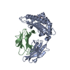 3rwcC 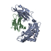 3rwdC  3rwfC  3rwgC 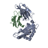 3rwhC  3rwiC 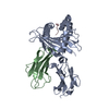 3rwjC  2bvoS C: citing same article ( S: Starting model for refinement |
|---|---|
| Similar structure data |


-Links
 PDBj
PDBj

- Assembly
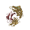
| Deposited unit | 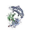
| ||||||||
|---|---|---|---|---|---|---|---|---|---|
| 1 |
| ||||||||
| Unit cell |
|
-Components
| #1: Protein | MHC class I Mass: 32025.082 Da / Num. of mol.: 1 / Fragment: residues 24-297 Source method: isolated from a genetically manipulated source Source: (gene. exp.) Macaca mulatta (Rhesus monkey) / Gene: MHCI-B / Production host:  Escherichia coli (E. coli) / References: UniProt: Q9GJ77 Escherichia coli (E. coli) / References: UniProt: Q9GJ77 | ||
|---|---|---|---|
| #2: Protein | Beta-2 microglobulin Mass: 11660.079 Da / Num. of mol.: 1 Source method: isolated from a genetically manipulated source Source: (gene. exp.) Macaca mulatta (Rhesus monkey) / Gene: B2M / Production host: Escherichia coli (E. coli) / References: UniProt: Q6V7J5 | ||
| #3: Protein/peptide | Mass: 1259.430 Da / Num. of mol.: 1 / Source method: obtained synthetically / Details: synthtic peptide / Source: (synth.) Simian immunodeficiency virus / References: UniProt: Q5QGH9 | ||
| #4: Chemical | Diethylene glycol  Mass: 106.120 Da / Num. of mol.: 2 / Source method: obtained synthetically / Formula: C4H10O3 Mass: 106.120 Da / Num. of mol.: 2 / Source method: obtained synthetically / Formula: C4H10O3#5: Water | ChemComp-HOH / | Water Mass: 18.015 Da / Num. of mol.: 268 / Source method: isolated from a natural source / Formula: H2O Mass: 18.015 Da / Num. of mol.: 268 / Source method: isolated from a natural source / Formula: H2O |
-Experimental details
-Experiment
| Experiment | Method: X-RAY DIFFRACTION / Number of used crystals: 1 |
|---|
- Sample preparation
| Crystal | Density Matthews: 2.83 Å3/Da / Density % sol: 56.48 % |
|---|
-Data collection
| Diffraction source | Source: ROTATING ANODE / Type: RIGAKU |
|---|---|
| Detector | Type: RIGAKU RAXIS IV++ / Detector: IMAGE PLATE / Date: Jun 18, 2009 |
| Radiation | Protocol: SINGLE WAVELENGTH / Monochromatic (M) / Laue (L): M / Scattering type: x-ray |
| Radiation wavelength | Relative weight: 1 |
| Reflection | Resolution: 2.35→50 Å / Num. obs: 20992 / Biso Wilson estimate: 37.66 Å2 |
- Processing
| Software | Name: PHENIX / Version: (phenix.refine: 1.6.4_486) / Classification: refinement | ||||||||||||||||||||||||||||||||||||||||||||||||||||||||
|---|---|---|---|---|---|---|---|---|---|---|---|---|---|---|---|---|---|---|---|---|---|---|---|---|---|---|---|---|---|---|---|---|---|---|---|---|---|---|---|---|---|---|---|---|---|---|---|---|---|---|---|---|---|---|---|---|---|
| Refinement | Method to determine structure: MOLECULAR REPLACEMENT Starting model: 2BVO Resolution: 2.4→28.55 Å / Occupancy max: 1 / Occupancy min: 0.46 / FOM work R set: 0.7987 / SU ML: 0.4 / σ(F): 1.34 / Phase error: 26.61 / Stereochemistry target values: ML
| ||||||||||||||||||||||||||||||||||||||||||||||||||||||||
| Solvent computation | Shrinkage radii: 0.9 Å / VDW probe radii: 1.11 Å / Solvent model: FLAT BULK SOLVENT MODEL / Bsol: 35.488 Å2 / ksol: 0.31 e/Å3 | ||||||||||||||||||||||||||||||||||||||||||||||||||||||||
| Displacement parameters | Biso max: 121.3 Å2 / Biso mean: 39.0923 Å2 / Biso min: 11.45 Å2
| ||||||||||||||||||||||||||||||||||||||||||||||||||||||||
| Refinement step | Cycle: LAST / Resolution: 2.4→28.55 Å
| ||||||||||||||||||||||||||||||||||||||||||||||||||||||||
| Refine LS restraints |
| ||||||||||||||||||||||||||||||||||||||||||||||||||||||||
| LS refinement shell | Refine-ID: X-RAY DIFFRACTION / Total num. of bins used: 7
| ||||||||||||||||||||||||||||||||||||||||||||||||||||||||
| Refinement TLS params. | Method: refined / Origin x: 26.0119 Å / Origin y: -0.4012 Å / Origin z: 20.11 Å
| ||||||||||||||||||||||||||||||||||||||||||||||||||||||||
| Refinement TLS group |
|
