 Movie
Movie Controller

[English] 日本語
 Yorodumi
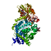
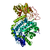
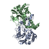
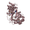
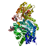
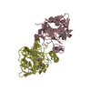
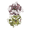
- PDB-3rrx: Crystal Structure of Q683A mutant of Exo-1,3/1,4-beta-glucanase (... -
+ Open data

- Basic information
| Entry | Database: PDB / ID: 3rrx | ||||||
|---|---|---|---|---|---|---|---|
| Title | Crystal Structure of Q683A mutant of Exo-1,3/1,4-beta-glucanase (ExoP) from Pseudoalteromonas sp. BB1 | ||||||
 Components Components | Exo-1,3/1,4-beta-glucanase | ||||||
 Keywords Keywords |  HYDROLASE / (ALPHA/BETA)8 BARREL / (ALPHA/BETA)6 SHEET HYDROLASE / (ALPHA/BETA)8 BARREL / (ALPHA/BETA)6 SHEET | ||||||
| Function / homology |  Function and homology informationbeta-glucosidase / hydrolase activity, hydrolyzing O-glycosyl compounds / carbohydrate metabolic process / metal ion binding Function and homology informationbeta-glucosidase / hydrolase activity, hydrolyzing O-glycosyl compounds / carbohydrate metabolic process / metal ion bindingSimilarity search - Function | ||||||
| Biological species |  Pseudoalteromonas sp. (bacteria) Pseudoalteromonas sp. (bacteria) | ||||||
| Method | X-RAY DIFFRACTION / SYNCHROTRON / MOLECULAR REPLACEMENT / Resolution: 1.9 Å | ||||||
 Authors Authors | Nakatani, Y. / Cutfield, S.M. / Cutfield, J.F. | ||||||
 Citation Citation | Journal: Febs J. / Year: 2012 Title: Structure and activity of exo-1,3/1,4-beta-glucanase from marine bacterium Pseudoalteromonas sp. BB1 showing a novel C-terminal domain Authors: Nakatani, Y. / Cutfield, S.M. / Cowieson, N.P. / Cutfield, J.F. #1: Journal: Appl.Environ.Microbiol. / Year: 2010 Title: Discovery and characterization of a distinctive exo-1,3/1,4-{beta}-glucanase from the marine bacterium Pseudoalteromonas sp. strain BB1 Authors: Nakatani, Y. / Lamont, I.L. / Cutfield, J.F. | ||||||
| History |
|
- Structure visualization
| Structure viewer | Molecule: MolmilJmol/JSmol |
|---|
- Downloads & links
-Download
| PDBx/mmCIF format | 3rrx.cif.gz | 153.2 KB | Display | PDBx/mmCIF format |
|---|---|---|---|---|
| PDB format | pdb3rrx.ent.gz | 114.5 KB | Display | PDB format |
| PDBx/mmJSON format | 3rrx.json.gz | Tree view | PDBx/mmJSON format | |
| Others |  Other downloads Other downloads |
-Validation report
| Arichive directory | https://data.pdbj.org/pub/pdb/validation_reports/rr/3rrxftp://data.pdbj.org/pub/pdb/validation_reports/rr/3rrx | HTTPS FTP |
|---|
-Related structure data
| Related structure data |  3f95C  3uszSC  3ut0C S: Starting model for refinement C: citing same article ( |
|---|---|
| Similar structure data |


-Links
 PDBj
PDBj- Assembly
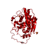
| Deposited unit | 
| ||||||||
|---|---|---|---|---|---|---|---|---|---|
| 1 |
| ||||||||
| Unit cell |
| ||||||||
| Components on special symmetry positions |
|
-Components
| #1: Protein | Mass: 90564.523 Da / Num. of mol.: 1 / Fragment: UNP RESIDUES 28-840 / Mutation: Q683A Source method: isolated from a genetically manipulated source Source: (gene. exp.) Pseudoalteromonas sp. (bacteria) / Strain: BB1 / Gene: exoP / Plasmid: PET21(D) / Production host: Escherichia coli (E. coli) / Strain (production host): BL21 (DE3) PLYSSReferences: UniProt: Q0QJA3, Hydrolases; Glycosylases; Glycosidases, i.e. enzymes that hydrolyse O- and S-glycosyl compounds | ||
|---|---|---|---|
| #2: Chemical | ChemComp-CA /   Mass: 40.078 Da / Num. of mol.: 1 / Source method: obtained synthetically / Formula: Ca Mass: 40.078 Da / Num. of mol.: 1 / Source method: obtained synthetically / Formula: Ca | ||
| #3: Chemical | ChemComp-NA /   Mass: 22.990 Da / Num. of mol.: 1 / Source method: obtained synthetically / Formula: Na Mass: 22.990 Da / Num. of mol.: 1 / Source method: obtained synthetically / Formula: Na | ||
| #4: Chemical | Ethylene glycol  Mass: 62.068 Da / Num. of mol.: 2 / Source method: obtained synthetically / Formula: C2H6O2 Mass: 62.068 Da / Num. of mol.: 2 / Source method: obtained synthetically / Formula: C2H6O2#5: Water | ChemComp-HOH / | Water Mass: 18.015 Da / Num. of mol.: 614 / Source method: isolated from a natural source / Formula: H2O Mass: 18.015 Da / Num. of mol.: 614 / Source method: isolated from a natural source / Formula: H2O |
-Experimental details
-Experiment
| Experiment | Method: X-RAY DIFFRACTION / Number of used crystals: 1 |
|---|
- Sample preparation
| Crystal | Density Matthews: 2.96 Å3/Da / Density % sol: 58.47 % |
|---|---|
| Crystal grow | Temperature: 291 K / Method: vapor diffusion, hanging drop / pH: 6.5 Details: 0.1M Bis-Tris propane pH 6.5, 20% PEG 3350, 0.2M sodium sulphate, VAPOR DIFFUSION, HANGING DROP, temperature 291K |
-Data collection
| Diffraction | Mean temperature: 113 K |
|---|---|
| Diffraction source | Source: SYNCHROTRON / Site: Australian Synchrotron  / Beamline: MX1 / Wavelength: 0.954 Å / Beamline: MX1 / Wavelength: 0.954 Å |
| Detector | Type: ADSC QUANTUM 210r / Detector: CCD / Date: Nov 16, 2010 |
| Radiation | Monochromator: mirror / Protocol: SINGLE WAVELENGTH / Monochromatic (M) / Laue (L): M / Scattering type: x-ray |
| Radiation wavelength | Wavelength: 0.954 Å / Relative weight: 1 |
| Reflection | Resolution: 1.9→19.98 Å / Num. obs: 84347 / % possible obs: 99.4 % / Redundancy: 4.3 % / Biso Wilson estimate: 21.5 Å2 / Rmerge(I) obs: 0.067 / Rsym value: 0.067 / Net I/σ(I): 15.2 |
| Reflection shell | Resolution: 1.9→2 Å / Redundancy: 4.3 % / Rmerge(I) obs: 0.45 / Mean I/σ(I) obs: 3 / Num. unique all: 12277 / Rsym value: 0.45 / % possible all: 100 |
- Processing
| Software |
| ||||||||||||||||||||||||||||||||||||||||||||||||||||||||||||||||||
|---|---|---|---|---|---|---|---|---|---|---|---|---|---|---|---|---|---|---|---|---|---|---|---|---|---|---|---|---|---|---|---|---|---|---|---|---|---|---|---|---|---|---|---|---|---|---|---|---|---|---|---|---|---|---|---|---|---|---|---|---|---|---|---|---|---|---|---|
| Refinement | Method to determine structure: MOLECULAR REPLACEMENT Starting model: PDB ENTRY 3USZ Resolution: 1.9→19.98 Å / FOM work R set: 0.8892 / SU ML: 0.18 / σ(F): 1.34 / Phase error: 18.26 / Stereochemistry target values: ML
| ||||||||||||||||||||||||||||||||||||||||||||||||||||||||||||||||||
| Solvent computation | Shrinkage radii: 0.9 Å / VDW probe radii: 1.11 Å / Solvent model: FLAT BULK SOLVENT MODEL / Bsol: 49.32 Å2 / ksol: 0.411 e/Å3 | ||||||||||||||||||||||||||||||||||||||||||||||||||||||||||||||||||
| Displacement parameters | Biso mean: 19.987 Å2
| ||||||||||||||||||||||||||||||||||||||||||||||||||||||||||||||||||
| Refinement step | Cycle: LAST / Resolution: 1.9→19.98 Å
| ||||||||||||||||||||||||||||||||||||||||||||||||||||||||||||||||||
| Refine LS restraints |
| ||||||||||||||||||||||||||||||||||||||||||||||||||||||||||||||||||
| LS refinement shell | Refine-ID: X-RAY DIFFRACTION / Total num. of bins used: 10
|
