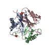
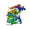
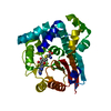
 Movie
Movie Controller


+ Open data

- Basic information
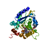
| Entry | Database: PDB / ID: 3rrw | ||||||
|---|---|---|---|---|---|---|---|
| Title | Crystal structure of the TL29 protein from Arabidopsis thaliana | ||||||
 Components Components | Thylakoid lumenal 29 kDa protein, chloroplastic | ||||||
 Keywords Keywords |  PLANT PROTEIN / chloroplast thylakoid lumen PLANT PROTEIN / chloroplast thylakoid lumen | ||||||
| Function / homology |  Function and homology informationthylakoid lumen / chloroplast thylakoid / thylakoid / chloroplast thylakoid lumen / Oxidoreductases / chloroplast thylakoid membrane / chloroplast / response to reactive oxygen species / hydrogen peroxide catabolic process / peroxidase activity ...thylakoid lumen / chloroplast thylakoid / thylakoid / chloroplast thylakoid lumen / Oxidoreductases / chloroplast thylakoid membrane / chloroplast / response to reactive oxygen species / hydrogen peroxide catabolic process / peroxidase activity / cellular response to oxidative stress / heme binding / nucleus / cytosol / cytoplasm Function and homology informationthylakoid lumen / chloroplast thylakoid / thylakoid / chloroplast thylakoid lumen / Oxidoreductases / chloroplast thylakoid membrane / chloroplast / response to reactive oxygen species / hydrogen peroxide catabolic process / peroxidase activity ...thylakoid lumen / chloroplast thylakoid / thylakoid / chloroplast thylakoid lumen / Oxidoreductases / chloroplast thylakoid membrane / chloroplast / response to reactive oxygen species / hydrogen peroxide catabolic process / peroxidase activity / cellular response to oxidative stress / heme binding / nucleus / cytosol / cytoplasmSimilarity search - Function | ||||||
| Biological species |  Arabidopsis thaliana (thale cress) Arabidopsis thaliana (thale cress) | ||||||
| Method | X-RAY DIFFRACTION / SYNCHROTRON / SAD / Resolution: 2.5 Å | ||||||
 Authors Authors | Lundberg, E. / Storm, P. / Schroder, W.P. / Funk, C. | ||||||
 Citation Citation | Journal: J.Struct.Biol. / Year: 2011 Title: Crystal structure of the TL29 protein from Arabidopsis thaliana: An APX homolog without peroxidase activity. Authors: Lundberg, E. / Storm, P. / Schroder, W.P. / Funk, C. | ||||||
| History |
|
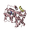
- Structure visualization
| Structure viewer | Molecule: MolmilJmol/JSmol |
|---|
- Downloads & links
-Download
| PDBx/mmCIF format | 3rrw.cif.gz | 203.5 KB | Display | PDBx/mmCIF format |
|---|---|---|---|---|
| PDB format | pdb3rrw.ent.gz | 172.6 KB | Display | PDB format |
| PDBx/mmJSON format | 3rrw.json.gz | Tree view | PDBx/mmJSON format | |
| Others |  Other downloads Other downloads |
-Validation report
| Arichive directory | https://data.pdbj.org/pub/pdb/validation_reports/rr/3rrwftp://data.pdbj.org/pub/pdb/validation_reports/rr/3rrw | HTTPS FTP |
|---|
-Related structure data
| Similar structure data |
|---|


-Links
 PDBj
PDBj
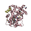
- Assembly
| Deposited unit | 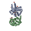
| ||||||||
|---|---|---|---|---|---|---|---|---|---|
| 1 | 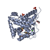
| ||||||||
| 2 | 
| ||||||||
| Unit cell |
|
-Components
| #1: Protein | Mass: 29647.926 Da / Num. of mol.: 2 / Fragment: Residues 83-349 Source method: isolated from a genetically manipulated source Source: (gene. exp.) Arabidopsis thaliana (thale cress) / Gene: TL29, APX4, At4g09010, F23J3.40 / Plasmid: pET-46 Ek/LIC / Production host:  Escherichia coli (E. coli) / Strain (production host): Rosetta2 / References: UniProt: P82281 Escherichia coli (E. coli) / Strain (production host): Rosetta2 / References: UniProt: P82281#2: Chemical | Phosphate  Mass: 94.971 Da / Num. of mol.: 2 / Source method: obtained synthetically / Formula: PO4 Mass: 94.971 Da / Num. of mol.: 2 / Source method: obtained synthetically / Formula: PO4#3: Chemical |   Mass: 40.078 Da / Num. of mol.: 2 / Source method: obtained synthetically / Formula: Ca Mass: 40.078 Da / Num. of mol.: 2 / Source method: obtained synthetically / Formula: Ca#4: Chemical | ChemComp-GOL / Glycerol  Mass: 92.094 Da / Num. of mol.: 5 / Source method: obtained synthetically / Formula: C3H8O3 Mass: 92.094 Da / Num. of mol.: 5 / Source method: obtained synthetically / Formula: C3H8O3#5: Water | ChemComp-HOH / | Water Mass: 18.015 Da / Num. of mol.: 43 / Source method: isolated from a natural source / Formula: H2O Mass: 18.015 Da / Num. of mol.: 43 / Source method: isolated from a natural source / Formula: H2O |
|---|
-Experimental details
-Experiment
| Experiment | Method: X-RAY DIFFRACTION / Number of used crystals: 1 |
|---|
- Sample preparation
| Crystal | Density Matthews: 2.58 Å3/Da / Density % sol: 52.31 % |
|---|---|
| Crystal grow | Temperature: 291 K / Method: vapor diffusion, hanging drop / pH: 4.8 Details: Reservoir solution: 16 % PEG 4000, 0.2M potassium phosphate, 10 % glycerol. Protein solution: 16 mM sodium phosphate, 0.1 M NaCl, 10 % glycerol, pH 4.8, VAPOR DIFFUSION, HANGING DROP, temperature 291K |
-Data collection
| Diffraction | Mean temperature: 100 K |
|---|---|
| Diffraction source | Source: SYNCHROTRON / Site: MAX II  / Beamline: I911-3 / Wavelength: 0.97926 Å / Beamline: I911-3 / Wavelength: 0.97926 Å |
| Detector | Type: MARMOSAIC 225 mm CCD / Detector: CCD / Date: May 6, 2009 |
| Radiation | Monochromator: SI (111) / Protocol: SINGLE WAVELENGTH / Monochromatic (M) / Laue (L): M / Scattering type: x-ray |
| Radiation wavelength | Wavelength: 0.97926 Å / Relative weight: 1 |
| Reflection | Resolution: 2.5→48.28 Å / Num. all: 22173 / Num. obs: 22173 / % possible obs: 99.9 % / Observed criterion σ(F): 2 / Observed criterion σ(I): 2 / Redundancy: 7.2 % / Rmerge(I) obs: 0.092 |
| Reflection shell | Resolution: 2.5→2.64 Å / Redundancy: 7.3 % / Rmerge(I) obs: 0.346 / Mean I/σ(I) obs: 4.3 / Num. unique all: 3160 / % possible all: 100 |
- Processing
| Software |
| |||||||||||||||||||||||||||||||||||||||||||||||||||||||||||||||||||||||||||
|---|---|---|---|---|---|---|---|---|---|---|---|---|---|---|---|---|---|---|---|---|---|---|---|---|---|---|---|---|---|---|---|---|---|---|---|---|---|---|---|---|---|---|---|---|---|---|---|---|---|---|---|---|---|---|---|---|---|---|---|---|---|---|---|---|---|---|---|---|---|---|---|---|---|---|---|---|
| Refinement | Method to determine structure: SAD / Resolution: 2.5→46.17 Å / Cor.coef. Fo:Fc: 0.93 / Cor.coef. Fo:Fc free: 0.901 / SU B: 21.384 / SU ML: 0.223 / Cross valid method: THROUGHOUT / ESU R Free: 0.306 / Stereochemistry target values: MAXIMUM LIKELIHOOD / Details: HYDROGENS HAVE BEEN ADDED IN THE RIDING POSITIONS
| |||||||||||||||||||||||||||||||||||||||||||||||||||||||||||||||||||||||||||
| Solvent computation | Ion probe radii: 0.8 Å / Shrinkage radii: 0.8 Å / VDW probe radii: 1.4 Å / Solvent model: MASK | |||||||||||||||||||||||||||||||||||||||||||||||||||||||||||||||||||||||||||
| Displacement parameters | Biso mean: 50.468 Å2
| |||||||||||||||||||||||||||||||||||||||||||||||||||||||||||||||||||||||||||
| Refinement step | Cycle: LAST / Resolution: 2.5→46.17 Å
| |||||||||||||||||||||||||||||||||||||||||||||||||||||||||||||||||||||||||||
| Refine LS restraints |
| |||||||||||||||||||||||||||||||||||||||||||||||||||||||||||||||||||||||||||
| LS refinement shell | Resolution: 2.5→2.565 Å / Total num. of bins used: 20
| |||||||||||||||||||||||||||||||||||||||||||||||||||||||||||||||||||||||||||
| Refinement TLS params. | Method: refined / Refine-ID: X-RAY DIFFRACTION
| |||||||||||||||||||||||||||||||||||||||||||||||||||||||||||||||||||||||||||
| Refinement TLS group |
|
