 Movie
Movie Controller


+ Open data

- Basic information
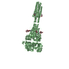
| Entry | Database: PDB / ID: 3rrr | ||||||
|---|---|---|---|---|---|---|---|
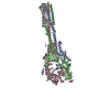
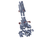
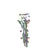
| Title | Structure of the RSV F protein in the post-fusion conformation | ||||||
 Components Components | (Fusion glycoprotein F0) x 2 | ||||||
 Keywords Keywords |  VIRAL PROTEIN / six-helix bundle / membrane fusion VIRAL PROTEIN / six-helix bundle / membrane fusion | ||||||
| Function / homology |  Function and homology information Function and homology informationpositive regulation of syncytium formation by virus / host cell Golgi membrane / entry receptor-mediated virion attachment to host cell / symbiont entry into host cell / fusion of virus membrane with host plasma membrane / viral envelope / host cell plasma membrane / virion membrane / membrane / identical protein binding / plasma membraneSimilarity search - Function | ||||||
| Biological species |  Human respiratory syncytial virus Human respiratory syncytial virus | ||||||
| Method | X-RAY DIFFRACTION / SYNCHROTRON / MOLECULAR REPLACEMENT / Resolution: 2.821 Å | ||||||
 Authors Authors | McLellan, J.S. / Yongping, Y. / Graham, B.S. / Kwong, P.D. | ||||||
 Citation Citation | Journal: J.Virol. / Year: 2011 Title: Structure of respiratory syncytial virus fusion glycoprotein in the postfusion conformation reveals preservation of neutralizing epitopes. Authors: McLellan, J.S. / Yang, Y. / Graham, B.S. / Kwong, P.D. | ||||||
| History |
|
- Structure visualization
| Structure viewer | Molecule: MolmilJmol/JSmol |
|---|
- Downloads & links
-Download
| PDBx/mmCIF format | 3rrr.cif.gz | 1.4 MB | Display | PDBx/mmCIF format |
|---|---|---|---|---|
| PDB format | pdb3rrr.ent.gz | 1.2 MB | Display | PDB format |
| PDBx/mmJSON format | 3rrr.json.gz | Tree view | PDBx/mmJSON format | |
| Others |  Other downloads Other downloads |
-Validation report
| Arichive directory | https://data.pdbj.org/pub/pdb/validation_reports/rr/3rrrftp://data.pdbj.org/pub/pdb/validation_reports/rr/3rrr | HTTPS FTP |
|---|
-Related structure data
| Related structure data |  3rrtC 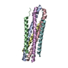 1g2cS C: citing same article ( S: Starting model for refinement |
|---|---|
| Similar structure data |


-Links
 PDBj
PDBj

- Assembly
| Deposited unit | 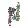
| ||||||||
|---|---|---|---|---|---|---|---|---|---|
| 1 | 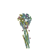
| ||||||||
| 2 | 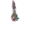
| ||||||||
| Unit cell |
|
-Components
| #1: Protein | Mass: 9529.798 Da / Num. of mol.: 6 / Fragment: unp residues 26-109 Source method: isolated from a genetically manipulated source Source: (gene. exp.) Human respiratory syncytial virus / Plasmid: paH / Cell line (production host): HEK293F / Production host:  homo sapiens (human) / References: UniProt: Q84850 homo sapiens (human) / References: UniProt: Q84850#2: Protein | Mass: 41323.137 Da / Num. of mol.: 6 / Fragment: unp residues 147-513 Source method: isolated from a genetically manipulated source Source: (gene. exp.) Human respiratory syncytial virus / Plasmid: paH / Cell line (production host): HEK293F / Production host: homo sapiens (human) / References: UniProt: Q84850, UniProt: P03420*PLUS#3: Sugar | ChemComp-NAG / N-Acetylglucosamine  Type: D-saccharide, beta linking / Mass: 221.208 Da / Num. of mol.: 12 Type: D-saccharide, beta linking / Mass: 221.208 Da / Num. of mol.: 12Source method: isolated from a genetically manipulated source Formula: C8H15NO6 Sequence details | 1) THE CRYSTALLIZED SEQUENCE CORRESPONDS TO A VIRAL STRAIN NOT PRESENT IN THE DATABASE UNP ENTRY ...1) THE CRYSTALLIZ | |
|---|
-Experimental details
-Experiment
| Experiment | Method: X-RAY DIFFRACTION / Number of used crystals: 1 |
|---|
- Sample preparation
| Crystal | Density Matthews: 3.9 Å3/Da / Density % sol: 68.47 % |
|---|---|
| Crystal grow | Temperature: 293 K / Method: vapor diffusion, hanging drop / pH: 5.5 Details: 20% (w/v) PEG 3000, 0.1 M sodium citrate, pH 5.5, VAPOR DIFFUSION, HANGING DROP, temperature 293K |
-Data collection
| Diffraction | Mean temperature: 200 K |
|---|---|
| Diffraction source | Source: SYNCHROTRON / Site: APS  / Beamline: 22-ID / Wavelength: 1 Å / Beamline: 22-ID / Wavelength: 1 Å |
| Detector | Type: MARMOSAIC 300 mm CCD / Detector: CCD / Date: Dec 20, 2010 |
| Radiation | Monochromator: Si(111) / Protocol: SINGLE WAVELENGTH / Monochromatic (M) / Laue (L): M / Scattering type: x-ray |
| Radiation wavelength | Wavelength: 1 Å / Relative weight: 1 |
| Reflection | Resolution: 2.8→50 Å / Num. all: 114379 / Num. obs: 76177 / % possible obs: 66.6 % / Observed criterion σ(F): 1 / Observed criterion σ(I): 1 / Redundancy: 3.2 % / Rmerge(I) obs: 0.159 / Rsym value: 0.159 / Net I/σ(I): 7.5 |
| Reflection shell | Resolution: 2.8→2.9 Å / Redundancy: 1.5 % / Rmerge(I) obs: 0.353 / Mean I/σ(I) obs: 1.4 / Num. unique all: 1902 / % possible all: 16.7 |
- Processing
| Software |
| |||||||||||||||||||||||||||||||||||||||||||||||||||||||||||||||||||||||||||||||||||||||||||||||||||||||||||||||||||||||||||||||||||||||||||||||||||||||||||||||||||||||||||||||||||||||||||||
|---|---|---|---|---|---|---|---|---|---|---|---|---|---|---|---|---|---|---|---|---|---|---|---|---|---|---|---|---|---|---|---|---|---|---|---|---|---|---|---|---|---|---|---|---|---|---|---|---|---|---|---|---|---|---|---|---|---|---|---|---|---|---|---|---|---|---|---|---|---|---|---|---|---|---|---|---|---|---|---|---|---|---|---|---|---|---|---|---|---|---|---|---|---|---|---|---|---|---|---|---|---|---|---|---|---|---|---|---|---|---|---|---|---|---|---|---|---|---|---|---|---|---|---|---|---|---|---|---|---|---|---|---|---|---|---|---|---|---|---|---|---|---|---|---|---|---|---|---|---|---|---|---|---|---|---|---|---|---|---|---|---|---|---|---|---|---|---|---|---|---|---|---|---|---|---|---|---|---|---|---|---|---|---|---|---|---|---|---|---|---|
| Refinement | Method to determine structure: MOLECULAR REPLACEMENT Starting model: pdb entry 1G2C Resolution: 2.821→44.209 Å / SU ML: 0.38 / σ(F): 0 / Phase error: 24.56 / Stereochemistry target values: ML
| |||||||||||||||||||||||||||||||||||||||||||||||||||||||||||||||||||||||||||||||||||||||||||||||||||||||||||||||||||||||||||||||||||||||||||||||||||||||||||||||||||||||||||||||||||||||||||||
| Solvent computation | Shrinkage radii: 0.17 Å / VDW probe radii: 0.4 Å / Solvent model: FLAT BULK SOLVENT MODEL / Bsol: 23.091 Å2 / ksol: 0.395 e/Å3 | |||||||||||||||||||||||||||||||||||||||||||||||||||||||||||||||||||||||||||||||||||||||||||||||||||||||||||||||||||||||||||||||||||||||||||||||||||||||||||||||||||||||||||||||||||||||||||||
| Displacement parameters |
| |||||||||||||||||||||||||||||||||||||||||||||||||||||||||||||||||||||||||||||||||||||||||||||||||||||||||||||||||||||||||||||||||||||||||||||||||||||||||||||||||||||||||||||||||||||||||||||
| Refinement step | Cycle: LAST / Resolution: 2.821→44.209 Å
| |||||||||||||||||||||||||||||||||||||||||||||||||||||||||||||||||||||||||||||||||||||||||||||||||||||||||||||||||||||||||||||||||||||||||||||||||||||||||||||||||||||||||||||||||||||||||||||
| Refine LS restraints |
| |||||||||||||||||||||||||||||||||||||||||||||||||||||||||||||||||||||||||||||||||||||||||||||||||||||||||||||||||||||||||||||||||||||||||||||||||||||||||||||||||||||||||||||||||||||||||||||
| LS refinement shell |
| |||||||||||||||||||||||||||||||||||||||||||||||||||||||||||||||||||||||||||||||||||||||||||||||||||||||||||||||||||||||||||||||||||||||||||||||||||||||||||||||||||||||||||||||||||||||||||||
| Refinement TLS params. | Method: refined / Refine-ID: X-RAY DIFFRACTION
| |||||||||||||||||||||||||||||||||||||||||||||||||||||||||||||||||||||||||||||||||||||||||||||||||||||||||||||||||||||||||||||||||||||||||||||||||||||||||||||||||||||||||||||||||||||||||||||
| Refinement TLS group |
|
