 Movie
Movie Controller


+ Open data

- Basic information
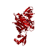
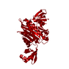
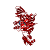
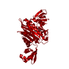
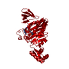
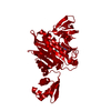
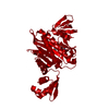
| Entry | Database: PDB / ID: 3rir | ||||||
|---|---|---|---|---|---|---|---|
| Title | Crystal Strucrture of Biotin Protein Ligase from S. aureus | ||||||
 Components Components | Biotin-[acetyl-CoA-carboxylase] ligase | ||||||
 Keywords Keywords |  LIGASE / Biotin protein ligase / Transcriptional repression / BCCP and DNA LIGASE / Biotin protein ligase / Transcriptional repression / BCCP and DNA | ||||||
| Function / homology |  Function and homology information Function and homology informationSH3 type barrels. - #100 / Bira Bifunctional Protein; Domain 2 / BirA Bifunctional Protein; domain 2 / Winged helix-like DNA-binding domain superfamily/Winged helix DNA-binding domain / SH3 type barrels. / Arc Repressor Mutant, subunit A / Roll / 2-Layer Sandwich / Orthogonal Bundle / Mainly Beta ...SH3 type barrels. - #100 / Bira Bifunctional Protein; Domain 2 / BirA Bifunctional Protein; domain 2 / Winged helix-like DNA-binding domain superfamily/Winged helix DNA-binding domain / SH3 type barrels. / Arc Repressor Mutant, subunit A / Roll / 2-Layer Sandwich / Orthogonal Bundle / Mainly Beta / Mainly Alpha / Alpha Beta Similarity search - Domain/homology | ||||||
| Biological species |   Staphylococcus aureus (bacteria) Staphylococcus aureus (bacteria) | ||||||
| Method | X-RAY DIFFRACTION / MOLECULAR REPLACEMENT / Resolution: 2.6 Å | ||||||
 Authors Authors | Wilce, M.C.J. | ||||||
 Citation Citation | Journal: To be published Title: Crystal Strucrture of Biotin Protein Ligase from S. aureus Authors: Pendini, N. / Yap, M. / Polyak, S. / Cowieson, N. / Daouda, T. / Booker, G. / Wallace, J. / Wilce, M.C.J. | ||||||
| History |
|
- Structure visualization
| Structure viewer | Molecule: MolmilJmol/JSmol |
|---|
- Downloads & links
-Download
| PDBx/mmCIF format | 3rir.cif.gz | 143.1 KB | Display | PDBx/mmCIF format |
|---|---|---|---|---|
| PDB format | pdb3rir.ent.gz | 112.1 KB | Display | PDB format |
| PDBx/mmJSON format | 3rir.json.gz | Tree view | PDBx/mmJSON format | |
| Others |  Other downloads Other downloads |
-Validation report
| Arichive directory | https://data.pdbj.org/pub/pdb/validation_reports/ri/3rirftp://data.pdbj.org/pub/pdb/validation_reports/ri/3rir | HTTPS FTP |
|---|
-Related structure data
| Related structure data |  1wq7S S: Starting model for refinement |
|---|---|
| Similar structure data |


-Links
 PDBj
PDBj- Assembly
| Deposited unit | 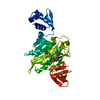
| ||||||||
|---|---|---|---|---|---|---|---|---|---|
| 1 | 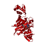
| ||||||||
| Unit cell |
|
-Components
| #1: Protein | Mass: 37186.910 Da / Num. of mol.: 1 Source method: isolated from a genetically manipulated source Source: (gene. exp.) Staphylococcus aureus (bacteria) / Strain: ECT-R 2 / Gene: ECTR2_1310 / Production host: Escherichia coli (E. coli)References: UniProt: E5R5T0, biotin-[biotin carboxyl-carrier protein] ligase |
|---|---|
| #2: Chemical | ChemComp-BT5 /   Type: RNA linking / Mass: 573.517 Da / Num. of mol.: 1 / Source method: obtained synthetically / Formula: C20H28N7O9PS Type: RNA linking / Mass: 573.517 Da / Num. of mol.: 1 / Source method: obtained synthetically / Formula: C20H28N7O9PS |
| #3: Water | ChemComp-HOH / Water Mass: 18.015 Da / Num. of mol.: 30 / Source method: isolated from a natural source / Formula: H2O Mass: 18.015 Da / Num. of mol.: 30 / Source method: isolated from a natural source / Formula: H2O |
-Experimental details
-Experiment
| Experiment | Method: X-RAY DIFFRACTION / Number of used crystals: 1 |
|---|
- Sample preparation
| Crystal | Density Matthews: 3.85 Å3/Da / Density % sol: 68.04 % |
|---|
-Data collection
| Diffraction | Mean temperature: 100 K |
|---|---|
| Diffraction source | Source: ROTATING ANODE / Type: RIGAKU MICROMAX-007 HF / Wavelength: 1.54 Å |
| Detector | Type: RIGAKU RAXIS IV++ / Detector: IMAGE PLATE |
| Radiation | Protocol: SINGLE WAVELENGTH / Monochromatic (M) / Laue (L): M / Scattering type: x-ray |
| Radiation wavelength | Wavelength: 1.54 Å / Relative weight: 1 |
| Reflection | Resolution: 2.6→35 Å / Num. obs: 18098 / % possible obs: 96.1 % / Observed criterion σ(F): 0 / Observed criterion σ(I): 0 / Redundancy: 3.8 % / Biso Wilson estimate: 35.2 Å2 / Rmerge(I) obs: 0.113 / Rsym value: 0.113 / Net I/σ(I): 7.1 |
- Processing
| Software |
| ||||||||||||||||||||||||||||||||||||||||||||||||||||||||||||||||||||||||||||||||||||||||||||||||||||
|---|---|---|---|---|---|---|---|---|---|---|---|---|---|---|---|---|---|---|---|---|---|---|---|---|---|---|---|---|---|---|---|---|---|---|---|---|---|---|---|---|---|---|---|---|---|---|---|---|---|---|---|---|---|---|---|---|---|---|---|---|---|---|---|---|---|---|---|---|---|---|---|---|---|---|---|---|---|---|---|---|---|---|---|---|---|---|---|---|---|---|---|---|---|---|---|---|---|---|---|---|---|
| Refinement | Method to determine structure: MOLECULAR REPLACEMENT Starting model: 1WQ7 Resolution: 2.6→19.925 Å / SU ML: 0.41 / Cross valid method: THROUGHOUT / σ(F): 1.34 / σ(I): 0 / Stereochemistry target values: ML
| ||||||||||||||||||||||||||||||||||||||||||||||||||||||||||||||||||||||||||||||||||||||||||||||||||||
| Solvent computation | Shrinkage radii: 0.77 Å / VDW probe radii: 0.9 Å / Solvent model: FLAT BULK SOLVENT MODEL / Bsol: 49.863 Å2 / ksol: 0.39 e/Å3 | ||||||||||||||||||||||||||||||||||||||||||||||||||||||||||||||||||||||||||||||||||||||||||||||||||||
| Displacement parameters | Biso mean: 35.1 Å2
| ||||||||||||||||||||||||||||||||||||||||||||||||||||||||||||||||||||||||||||||||||||||||||||||||||||
| Refinement step | Cycle: LAST / Resolution: 2.6→19.925 Å
| ||||||||||||||||||||||||||||||||||||||||||||||||||||||||||||||||||||||||||||||||||||||||||||||||||||
| Refine LS restraints |
| ||||||||||||||||||||||||||||||||||||||||||||||||||||||||||||||||||||||||||||||||||||||||||||||||||||
| LS refinement shell | Refine-ID: X-RAY DIFFRACTION / Total num. of bins used: 7
| ||||||||||||||||||||||||||||||||||||||||||||||||||||||||||||||||||||||||||||||||||||||||||||||||||||
| Refinement TLS params. | Method: refined / Refine-ID: X-RAY DIFFRACTION
| ||||||||||||||||||||||||||||||||||||||||||||||||||||||||||||||||||||||||||||||||||||||||||||||||||||
| Refinement TLS group |
|
