 Movie
Movie Controller

[English] 日本語
 Yorodumi
- PDB-3rho: Crystal structure of the E673Q MUTANT OF C-Terminal domain of 10'... -
+ Open data

- Basic information
| Entry | Database: PDB / ID: 3rho | ||||||
|---|---|---|---|---|---|---|---|
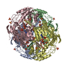
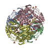
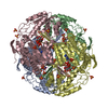
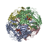
| Title | Crystal structure of the E673Q MUTANT OF C-Terminal domain of 10'FORMYLTETRAHYDROFOLATE DEHYDROGENASE in complex with NADP | ||||||
 Components Components | Aldehyde dehydrogenase 1 family, member L1 | ||||||
 Keywords Keywords |  OXIDOREDUCTASE / FDH OXIDOREDUCTASE / FDH | ||||||
| Function / homology |  Function and homology information Function and homology informationMetabolism of folate and pterines / aldehyde dehydrogenase (NADP+) activity / formyltetrahydrofolate dehydrogenase / formyltetrahydrofolate dehydrogenase activity / 10-formyltetrahydrofolate catabolic process / aldehyde dehydrogenase [NAD(P)+] activity / NADPH regeneration / aldehyde dehydrogenase (NAD+) activity / bile acid signaling pathway / folic acid metabolic process ...Metabolism of folate and pterines / aldehyde dehydrogenase (NADP+) activity / formyltetrahydrofolate dehydrogenase / formyltetrahydrofolate dehydrogenase activity / 10-formyltetrahydrofolate catabolic process / aldehyde dehydrogenase [NAD(P)+] activity / NADPH regeneration / aldehyde dehydrogenase (NAD+) activity / bile acid signaling pathway / folic acid metabolic process / tetrahydrofolate biosynthetic process / one-carbon metabolic process / protein-containing complex binding / protein-containing complex / cytosolSimilarity search - Function | ||||||
| Biological species |  Rattus norvegicus (Norway rat) Rattus norvegicus (Norway rat) | ||||||
| Method | X-RAY DIFFRACTION / SYNCHROTRON / MOLECULAR REPLACEMENT / Resolution: 2.26 Å | ||||||
 Authors Authors | Tsybovsky, Y. | ||||||
 Citation Citation | Journal: J.Biol.Chem. / Year: 2011 Title: Conserved catalytic residues of the ALDH1L1 aldehyde dehydrogenase domain control binding and discharging of the coenzyme. Authors: Tsybovsky, Y. / Krupenko, S.A. | ||||||
| History |
|
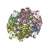
- Structure visualization
| Structure viewer | Molecule: MolmilJmol/JSmol |
|---|
- Downloads & links
-Download
| PDBx/mmCIF format | 3rho.cif.gz | 435.4 KB | Display | PDBx/mmCIF format |
|---|---|---|---|---|
| PDB format | pdb3rho.ent.gz | 355.8 KB | Display | PDB format |
| PDBx/mmJSON format | 3rho.json.gz | Tree view | PDBx/mmJSON format | |
| Others |  Other downloads Other downloads |
-Validation report
| Arichive directory | https://data.pdbj.org/pub/pdb/validation_reports/rh/3rhoftp://data.pdbj.org/pub/pdb/validation_reports/rh/3rho | HTTPS FTP |
|---|
-Related structure data
| Related structure data |  3rhjC  3rhlC  3rhmC  3rhpC  3rhqC  3rhrC  2o2pS C: citing same article ( S: Starting model for refinement |
|---|---|
| Similar structure data |


-Links
 PDBj
PDBj


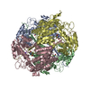
- Assembly
| Deposited unit | 
| ||||||||
|---|---|---|---|---|---|---|---|---|---|
| 1 |
| ||||||||
| Unit cell |
|
-Components
| #1: Protein | Mass: 56620.562 Da / Num. of mol.: 4 / Fragment: C-TERMINAL DOMAIN, RESIDUES 397-902 / Mutation: E673Q Source method: isolated from a genetically manipulated source Source: (gene. exp.) Rattus norvegicus (Norway rat) / Gene: Aldh1l1, Fthfd / Production host:  Escherichia coli (E. coli) / References: UniProt: Q5HZB2, UniProt: P28037*PLUS Escherichia coli (E. coli) / References: UniProt: Q5HZB2, UniProt: P28037*PLUS#2: Chemical | ChemComp-NAP / Nicotinamide adenine dinucleotide phosphate  Mass: 743.405 Da / Num. of mol.: 4 / Source method: obtained synthetically / Formula: C21H28N7O17P3 Mass: 743.405 Da / Num. of mol.: 4 / Source method: obtained synthetically / Formula: C21H28N7O17P3#3: Chemical | ChemComp-SO4 / Sulfate  Mass: 96.063 Da / Num. of mol.: 28 / Source method: obtained synthetically / Formula: SO4 Mass: 96.063 Da / Num. of mol.: 28 / Source method: obtained synthetically / Formula: SO4#4: Chemical | ChemComp-GOL / Glycerol  Mass: 92.094 Da / Num. of mol.: 4 / Source method: obtained synthetically / Formula: C3H8O3 Mass: 92.094 Da / Num. of mol.: 4 / Source method: obtained synthetically / Formula: C3H8O3#5: Water | ChemComp-HOH / | Water Mass: 18.015 Da / Num. of mol.: 1484 / Source method: isolated from a natural source / Formula: H2O Mass: 18.015 Da / Num. of mol.: 1484 / Source method: isolated from a natural source / Formula: H2O |
|---|
-Experimental details
-Experiment
| Experiment | Method: X-RAY DIFFRACTION / Number of used crystals: 1 |
|---|
- Sample preparation
| Crystal grow | Temperature: 293 K / Method: vapor diffusion, hanging drop / pH: 7.5 Details: 0.1M Tris pH 7.5, 1.6M Ammonium Sulfate, VAPOR DIFFUSION, HANGING DROP, temperature 293K |
|---|
-Data collection
| Diffraction | Mean temperature: 100 K |
|---|---|
| Diffraction source | Source: SYNCHROTRON / Site: APS  / Beamline: 22-ID / Wavelength: 1 Å / Beamline: 22-ID / Wavelength: 1 Å |
| Detector | Type: MAR scanner 300 mm plate / Detector: IMAGE PLATE / Date: Jul 21, 2007 |
| Radiation | Monochromator: double crystal / Protocol: SINGLE WAVELENGTH / Monochromatic (M) / Laue (L): M / Scattering type: x-ray |
| Radiation wavelength | Wavelength: 1 Å / Relative weight: 1 |
| Reflection | Resolution: 2.25→50 Å / Num. all: 213476 / Num. obs: 213476 / % possible obs: 99.9 % / Observed criterion σ(F): 0 / Observed criterion σ(I): 0 / Redundancy: 5.3 % / Rmerge(I) obs: 0.13 / Net I/σ(I): 9.7 |
| Reflection shell | Resolution: 2.25→2.33 Å / Redundancy: 4.9 % / Rmerge(I) obs: 0.547 / Mean I/σ(I) obs: 5.5 / % possible all: 99.9 |
- Processing
| Software |
| ||||||||||||||||||||||||||||||||||||||||||||||||||||||||||||||||||||||||||||||||||||||||||||||||||||||||||||||||||||||||||||||||||||||||||||||||||||||||||||||||||||||||||
|---|---|---|---|---|---|---|---|---|---|---|---|---|---|---|---|---|---|---|---|---|---|---|---|---|---|---|---|---|---|---|---|---|---|---|---|---|---|---|---|---|---|---|---|---|---|---|---|---|---|---|---|---|---|---|---|---|---|---|---|---|---|---|---|---|---|---|---|---|---|---|---|---|---|---|---|---|---|---|---|---|---|---|---|---|---|---|---|---|---|---|---|---|---|---|---|---|---|---|---|---|---|---|---|---|---|---|---|---|---|---|---|---|---|---|---|---|---|---|---|---|---|---|---|---|---|---|---|---|---|---|---|---|---|---|---|---|---|---|---|---|---|---|---|---|---|---|---|---|---|---|---|---|---|---|---|---|---|---|---|---|---|---|---|---|---|---|---|---|---|---|---|
| Refinement | Method to determine structure: MOLECULAR REPLACEMENT Starting model: PDB entry 2O2P Resolution: 2.26→49.15 Å / Cor.coef. Fo:Fc: 0.964 / Cor.coef. Fo:Fc free: 0.949 / SU B: 3.058 / SU ML: 0.077 / Cross valid method: THROUGHOUT / ESU R: 0.125 / ESU R Free: 0.12 / Stereochemistry target values: MAXIMUM LIKELIHOOD / Details: HYDROGENS HAVE BEEN ADDED IN THE RIDING POSITIONS
| ||||||||||||||||||||||||||||||||||||||||||||||||||||||||||||||||||||||||||||||||||||||||||||||||||||||||||||||||||||||||||||||||||||||||||||||||||||||||||||||||||||||||||
| Solvent computation | Ion probe radii: 0.8 Å / Shrinkage radii: 0.8 Å / VDW probe radii: 1.4 Å / Solvent model: MASK | ||||||||||||||||||||||||||||||||||||||||||||||||||||||||||||||||||||||||||||||||||||||||||||||||||||||||||||||||||||||||||||||||||||||||||||||||||||||||||||||||||||||||||
| Displacement parameters | Biso mean: 28.323 Å2
| ||||||||||||||||||||||||||||||||||||||||||||||||||||||||||||||||||||||||||||||||||||||||||||||||||||||||||||||||||||||||||||||||||||||||||||||||||||||||||||||||||||||||||
| Refinement step | Cycle: LAST / Resolution: 2.26→49.15 Å
| ||||||||||||||||||||||||||||||||||||||||||||||||||||||||||||||||||||||||||||||||||||||||||||||||||||||||||||||||||||||||||||||||||||||||||||||||||||||||||||||||||||||||||
| Refine LS restraints |
| ||||||||||||||||||||||||||||||||||||||||||||||||||||||||||||||||||||||||||||||||||||||||||||||||||||||||||||||||||||||||||||||||||||||||||||||||||||||||||||||||||||||||||
| LS refinement shell | Resolution: 2.26→2.313 Å / Total num. of bins used: 20
|
