 Movie
Movie Controller

[English] 日本語
 Yorodumi
- PDB-3rfi: Crystal structure of the saposin-like domain of plant aspartic pr... -
+ Open data

- Basic information
| Entry | Database: PDB / ID: 3rfi | ||||||
|---|---|---|---|---|---|---|---|
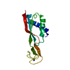
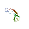
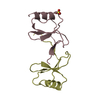
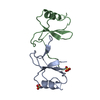
| Title | Crystal structure of the saposin-like domain of plant aspartic protease from Solanum tuberosum | ||||||
 Components Components | Asp | ||||||
 Keywords Keywords |  HYDROLASE / Aspartic protease / PSI / Saposin HYDROLASE / Aspartic protease / PSI / Saposin | ||||||
| Function / homology |  Function and homology information Function and homology information | ||||||
| Biological species |  Solanum tuberosum (potato) Solanum tuberosum (potato) | ||||||
| Method | X-RAY DIFFRACTION / MOLECULAR REPLACEMENT / Resolution: 1.9 Å | ||||||
 Authors Authors | Bhaumik, P. / Wlodawer, A. | ||||||
 Citation Citation | Journal: J.Biol.Chem. / Year: 2011 Title: Structure and Mechanism of the Saposin-like Domain of a Plant Aspartic Protease. Authors: Bryksa, B.C. / Bhaumik, P. / Magracheva, E. / De Moura, D.C. / Kurylowicz, M. / Zdanov, A. / Dutcher, J.R. / Wlodawer, A. / Yada, R.Y. | ||||||
| History |
|
- Structure visualization
| Structure viewer | Molecule: MolmilJmol/JSmol |
|---|
- Downloads & links
-Download
| PDBx/mmCIF format | 3rfi.cif.gz | 42.4 KB | Display | PDBx/mmCIF format |
|---|---|---|---|---|
| PDB format | pdb3rfi.ent.gz | 33.6 KB | Display | PDB format |
| PDBx/mmJSON format | 3rfi.json.gz | Tree view | PDBx/mmJSON format | |
| Others |  Other downloads Other downloads |
-Validation report
| Arichive directory | https://data.pdbj.org/pub/pdb/validation_reports/rf/3rfiftp://data.pdbj.org/pub/pdb/validation_reports/rf/3rfi | HTTPS FTP |
|---|
-Related structure data
| Similar structure data |
|---|


-Links
 PDBj
PDBj- Assembly
| Deposited unit | 
| ||||||||
|---|---|---|---|---|---|---|---|---|---|
| 1 |
| ||||||||
| 2 | 
| ||||||||
| Unit cell |
| ||||||||
| Components on special symmetry positions |
|
-Components
| #1: Protein | Mass: 11779.507 Da / Num. of mol.: 1 / Fragment: StAP_PSI (UNP Residues 301-403) Source method: isolated from a genetically manipulated source Source: (gene. exp.) Solanum tuberosum (potato) / Plasmid: pET32b(+) / Production host:  Escherichia coli (E. coli) / Strain (production host): Rosetta-gami B (DE3)pLysS / References: UniProt: Q6B9W9 Escherichia coli (E. coli) / Strain (production host): Rosetta-gami B (DE3)pLysS / References: UniProt: Q6B9W9 |
|---|---|
| #2: Water | ChemComp-HOH / Water Mass: 18.015 Da / Num. of mol.: 63 / Source method: isolated from a natural source / Formula: H2O Mass: 18.015 Da / Num. of mol.: 63 / Source method: isolated from a natural source / Formula: H2O |
-Experimental details
-Experiment
| Experiment | Method: X-RAY DIFFRACTION / Number of used crystals: 1 |
|---|
- Sample preparation
| Crystal | Density Matthews: 2.16 Å3/Da / Density % sol: 43.12 % |
|---|---|
| Crystal grow | Temperature: 293 K / Method: vapor diffusion, sitting drop Details: 0.2M Lithium sulfate monohydrate, 20% PEG 3350, VAPOR DIFFUSION, SITTING DROP, temperature 293K |
-Data collection
| Diffraction | Mean temperature: 100 K |
|---|---|
| Diffraction source | Source: ROTATING ANODE / Type: OTHER / Wavelength: 1.5418 Å |
| Detector | Type: MAR scanner 345 mm plate / Detector: IMAGE PLATE / Date: Nov 9, 2009 |
| Radiation | Protocol: SINGLE WAVELENGTH / Monochromatic (M) / Laue (L): M / Scattering type: x-ray |
| Radiation wavelength | Wavelength: 1.5418 Å / Relative weight: 1 |
| Reflection | Resolution: 1.9→40 Å / Num. all: 8363 / Num. obs: 8355 / % possible obs: 99.9 % / Redundancy: 10.3 % / Rmerge(I) obs: 0.059 / Net I/σ(I): 24.1 |
| Reflection shell | Resolution: 1.9→2 Å / Redundancy: 10.3 % / Rmerge(I) obs: 0.9 / Mean I/σ(I) obs: 2.87 / Num. unique all: 1159 / % possible all: 100 |
- Processing
| Software |
| ||||||||||||||||||||||||||||||||||||||||||||||||||||||||||||||||||||||||||||||||||||||||||||||||||||
|---|---|---|---|---|---|---|---|---|---|---|---|---|---|---|---|---|---|---|---|---|---|---|---|---|---|---|---|---|---|---|---|---|---|---|---|---|---|---|---|---|---|---|---|---|---|---|---|---|---|---|---|---|---|---|---|---|---|---|---|---|---|---|---|---|---|---|---|---|---|---|---|---|---|---|---|---|---|---|---|---|---|---|---|---|---|---|---|---|---|---|---|---|---|---|---|---|---|---|---|---|---|
| Refinement | Method to determine structure: MOLECULAR REPLACEMENT / Resolution: 1.9→25 Å / Cor.coef. Fo:Fc: 0.96 / Cor.coef. Fo:Fc free: 0.92 / SU B: 7.179 / SU ML: 0.094 / Cross valid method: THROUGHOUT / ESU R: 0.133 / ESU R Free: 0.143 / Stereochemistry target values: MAXIMUM LIKELIHOOD
| ||||||||||||||||||||||||||||||||||||||||||||||||||||||||||||||||||||||||||||||||||||||||||||||||||||
| Solvent computation | Ion probe radii: 0.8 Å / Shrinkage radii: 0.8 Å / VDW probe radii: 1.2 Å / Solvent model: MASK | ||||||||||||||||||||||||||||||||||||||||||||||||||||||||||||||||||||||||||||||||||||||||||||||||||||
| Displacement parameters | Biso mean: 18.949 Å2
| ||||||||||||||||||||||||||||||||||||||||||||||||||||||||||||||||||||||||||||||||||||||||||||||||||||
| Refinement step | Cycle: LAST / Resolution: 1.9→25 Å
| ||||||||||||||||||||||||||||||||||||||||||||||||||||||||||||||||||||||||||||||||||||||||||||||||||||
| Refine LS restraints |
| ||||||||||||||||||||||||||||||||||||||||||||||||||||||||||||||||||||||||||||||||||||||||||||||||||||
| LS refinement shell | Resolution: 1.9→1.949 Å / Total num. of bins used: 20
| ||||||||||||||||||||||||||||||||||||||||||||||||||||||||||||||||||||||||||||||||||||||||||||||||||||
| Refinement TLS params. | Method: refined / Refine-ID: X-RAY DIFFRACTION
| ||||||||||||||||||||||||||||||||||||||||||||||||||||||||||||||||||||||||||||||||||||||||||||||||||||
| Refinement TLS group |
|
