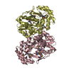
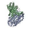
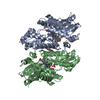
ANALYTICAL SIZE EXCLUSION CHROMATOGRAPHY WITH STATIC LIGHT SCATTERING SUPPORTS THE ASSIGNMENT OF A DIMER AS A SIGNIFICANT OLIGOMERIZATION STATE IN SOLUTION.
-
Components
-
Protein , 1 types, 1 molecules A
#1: Protein
Iron-containingalcoholdehydrogenase
Mass: 42629.930 Da / Num. of mol.: 1 Source method: isolated from a genetically manipulated source Source: (gene. exp.) Shewanella denitrificans (bacteria) / Strain: OS217 / ATCC BAA-1090 / DSM 15013 / Gene: Sden_2133 / Plasmid: SpeedET / Production host: Escherichia coli (E. coli) / Strain (production host): HK100 / References: UniProt: Q12MB1
Monochromator: double crystal monochromator / Protocol: SINGLE WAVELENGTH / Monochromatic (M) / Laue (L): M / Scattering type: x-ray
Radiation wavelength
Wavelength: 0.97936 Å / Relative weight: 1
Reflection
Resolution: 2.12→29.947 Å / Num. all: 58332 / Num. obs: 58332 / % possible obs: 100 % / Redundancy: 7.4 % / Biso Wilson estimate: 33.086 Å2 / Rsym value: 0.166 / Net I/σ(I): 8.1
Reflection shell
Diffraction-ID: 1
Resolution (Å)
Redundancy (%)
Rmerge(I) obs
Mean I/σ(I) obs
Num. measured all
Num. unique all
Rsym value
% possible all
2.12-2.18
7.5
1.388
1.6
31495
4226
1.388
100
2.18-2.23
7.5
1.079
2
31047
4157
1.079
100
2.23-2.3
7.5
0.859
2.4
30154
4047
0.859
100
2.3-2.37
7.5
0.729
2.8
29292
3925
0.729
100
2.37-2.45
7.5
0.604
3.2
28484
3814
0.604
100
2.45-2.53
7.5
0.473
4
27482
3678
0.473
100
2.53-2.63
7.5
0.418
4.5
26736
3575
0.418
100
2.63-2.74
7.5
0.328
5.4
25526
3415
0.328
100
2.74-2.86
7.4
0.259
6.5
24586
3301
0.259
100
2.86-3
7.5
0.209
7.9
23627
3160
0.209
100
3-3.16
7.5
0.167
9.6
22453
3013
0.167
100
3.16-3.35
7.5
0.136
11.5
21372
2867
0.136
100
3.35-3.58
7.4
0.114
13.8
19903
2682
0.114
100
3.58-3.87
7.4
0.098
16.6
18755
2528
0.098
100
3.87-4.24
7.4
0.09
18.2
17086
2312
0.09
100
4.24-4.74
7.3
0.079
20.1
15544
2118
0.079
100
4.74-5.47
7.3
0.081
19.3
13753
1886
0.081
100
5.47-6.7
7.2
0.094
17.6
11614
1615
0.094
100
6.7-9.48
7
0.081
19.8
8971
1280
0.081
100
9.48-29.947
6.4
0.066
20.7
4693
733
0.066
97
-
Phasing
Phasing
Method: SAD
-
Processing
Software
Name
Version
Classification
NB
MolProbity
3beta29
modelbuilding
PDB_EXTRACT
3.1
dataextraction
SHELX
phasing
SHARP
phasing
SCALA
3.3.15
datascaling
REFMAC
5.5.0110
refinement
MOSFLM
datareduction
SHELXD
phasing
Refinement
Method to determine structure: SAD / Resolution: 2.12→29.947 Å / Cor.coef. Fo:Fc: 0.958 / Cor.coef. Fo:Fc free: 0.955 / Occupancy max: 1 / Occupancy min: 0.35 / SU B: 5.271 / SU ML: 0.074 / Cross valid method: THROUGHOUT / σ(F): 0 / ESU R: 0.11 / ESU R Free: 0.104 Stereochemistry target values: MAXIMUM LIKELIHOOD WITH PHASES Details: 1. A MET-INHIBITION PROTOCOL WAS USED FOR SELENOMETHIONINE INCORPORATION DURING PROTEIN EXPRESSION. THE OCCUPANCY OF THE SE ATOMS IN THE MSE RESIDUES WAS REDUCED TO 0.75 FOR THE REDUCED ...Details: 1. A MET-INHIBITION PROTOCOL WAS USED FOR SELENOMETHIONINE INCORPORATION DURING PROTEIN EXPRESSION. THE OCCUPANCY OF THE SE ATOMS IN THE MSE RESIDUES WAS REDUCED TO 0.75 FOR THE REDUCED SCATTERING POWER DUE TO PARTIAL S-MET INCORPORATION. 2. HYDROGENS HAVE BEEN ADDED IN THE RIDING POSITIONS. 3. ATOM RECORD CONTAINS SUM OF TLS AND RESIDUAL B FACTORS. ANISOU RECORD CONTAINS SUM OF TLS AND RESIDUAL U FACTORS. 4. WATERS WERE EXCLUDED FROM AUTOMATIC TLS ASSIGNMENT. 5. TLS GROUPS WERE ASSIGNED WITH THE AID OF THE TLSMD (MOTION DETERMINATION) SERVER. 6. CALCIUM AND CHLORIDE IONS, AND HEPES (EPE) MOLECULES FROM THE CRYSTALLIZATION ARE MODELED INTO THE STRUCTURE. 7. X-RAY ANOMALOUS SCATTERING MEASUREMENTS INDICATE A MIXTURE OF AN IRON (FE) AND A NICKEL (NI) ATOM IS WITHIN COORDINATION DISTANCE OF THE SIDE CHAINS OF ASP-194, HIS-198, HIS-258, AND HIS-272. THE OCCUPANCIES OF THE METAL IONS WERE ESTIMATED FROM THE RATIOS OF THEIR ANOMALOUS DIFFERENCE MAP PEAK HEIGHTS AT WAVELENGTHS ABOVE AND BELOW THE FE AND NI K-SHELL ABSORPTION EDGES. REDUCING THE TOTAL OCCUPANCY OF THE FE/NI SITE TO 0.8 RESULTED IN BETTER AGREEMENT BETWEEN THE B-FACTORS OF THE METAL ATOMS AND ATOMS ON THE PROTEIN WITHIN COORDINATION DISTANCE. 8. ADDITIONAL ELECTRON DENSITY AT THE PUTATIVE ACTIVE SITE WAS MODELED AS AN UNKNOWN LIGAND (UNL). THE UNL COULD REPRESENT AN ANALOG TO THE SUBSTITUTED NADP THAT WAS IDENTIFIED IN THE E.COLI ALCOHOL DEHYDROGENASE YQHD (1OJ7) STRUCTURE. HOWEVER, MODELING IT AS AN NC5 AND NC6 SUBSTITUTED NAD RESULTED IN RESIDUAL DIFFERENCE DENSITY AT THE SITE SUGGESTING THAT EITHER THIS WAS NOT FULLY OCCUPIED AND WAS A MIXTURE OF COMPOUNDS OR IT WAS NOT THE CORRECT ANALOG. WITHOUT ADDITIONAL DATA TO IDENTIFY THE COMPOUND(S), WE HAVE CHOSEN TO MODEL IT AS A UNL ADJACENT TO THE NAD AND METAL SITE.
Rfactor
Num. reflection
% reflection
Selection details
Rfree
0.1968
2951
5.1 %
RANDOM
Rwork
0.1819
-
-
-
obs
0.1826
58283
99.92 %
-
Solvent computation
Ion probe radii: 0.8 Å / Shrinkage radii: 0.8 Å / VDW probe radii: 1.4 Å / Solvent model: MASK
In the structure databanks used in Yorodumi, some data are registered as the other names, "COVID-19 virus" and "2019-nCoV". Here are the details of the virus and the list of structure data.
Jan 31, 2019. EMDB accession codes are about to change! (news from PDBe EMDB page)
EMDB accession codes are about to change! (news from PDBe EMDB page)
The allocation of 4 digits for EMDB accession codes will soon come to an end. Whilst these codes will remain in use, new EMDB accession codes will include an additional digit and will expand incrementally as the available range of codes is exhausted. The current 4-digit format prefixed with “EMD-” (i.e. EMD-XXXX) will advance to a 5-digit format (i.e. EMD-XXXXX), and so on. It is currently estimated that the 4-digit codes will be depleted around Spring 2019, at which point the 5-digit format will come into force.
The EM Navigator/Yorodumi systems omit the EMD- prefix.
Related info.:Q: What is EMD? / ID/Accession-code notation in Yorodumi/EM Navigator
Yorodumi is a browser for structure data from EMDB, PDB, SASBDB, etc.
This page is also the successor to EM Navigator detail page, and also detail information page/front-end page for Omokage search.
The word "yorodu" (or yorozu) is an old Japanese word meaning "ten thousand". "mi" (miru) is to see.
Related info.:EMDB / PDB / SASBDB / Comparison of 3 databanks / Yorodumi Search / Aug 31, 2016. New EM Navigator & Yorodumi / Yorodumi Papers / Jmol/JSmol / Function and homology information / Changes in new EM Navigator and Yorodumi
 Movie
Movie Controller

 Yorodumi
 Open data
 Basic information
 Components
 Keywords
 OXIDOREDUCTASE /
  Function and homology information
 Authors
 Citation
 Structure visualization
 Downloads & links
 Other downloads
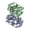


 PDBj
PDBj

 Assembly


 Mass: 663.425 Da / Num. of mol.: 1 / Source method: obtained synthetically / Formula: C21H27N7O14P2 / Comment: NAD*YM
 Mass: 238.305 Da / Num. of mol.: 1 / Source method: obtained synthetically / Formula: C8H18N2O4S / Comment: pH buffer*YM
 Mass: 55.845 Da / Num. of mol.: 1 / Source method: obtained synthetically / Formula: Fe
 Mass: 58.693 Da / Num. of mol.: 1 / Source method: obtained synthetically / Formula: Ni
 Mass: 35.453 Da / Num. of mol.: 4 / Source method: obtained synthetically / Formula: Cl
 Mass: 40.078 Da / Num. of mol.: 2 / Source method: obtained synthetically / Formula: Ca
 Mass: 106.120 Da / Num. of mol.: 2 / Source method: obtained synthetically / Formula: C4H10O3
 Sample preparation
 / Beamline: BL9-2 / Wavelength: 0.97936
  Processing