 Movie
Movie Controller


+ Open data

- Basic information
| Entry | Database: PDB / ID: 3rav | ||||||
|---|---|---|---|---|---|---|---|
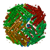
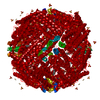
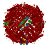
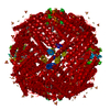
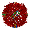
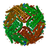
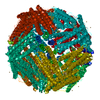
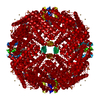
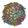
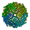
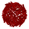
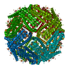
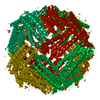
| Title | Horse spleen apo-ferritin with bound Pentobarbital | ||||||
 Components Components | Ferritin light chain | ||||||
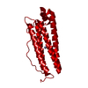
 Keywords Keywords | METAL BINDING PROTEIN / 4-HELIX BUNDLE / IRON / IRON STORAGE / METAL-BINDING | ||||||
| Function / homology |  Function and homology information Function and homology information: / intracellular sequestering of iron ion / ferric iron binding / ferrous iron binding / iron ion transport / iron ion binding / cytoplasmSimilarity search - Function | ||||||
| Biological species |  Equus caballus (horse) Equus caballus (horse) | ||||||
| Method | X-RAY DIFFRACTION / SYNCHROTRON / molecular replacement, molecular replacement / molecular replacement / Resolution: 1.9 Å | ||||||
 Authors Authors | Oakley, S.H. / Vedula, L.S. / Xi, J. / Liu, R. / Eckenhoff, R.G. / Loll, P.J. | ||||||
 Citation Citation | Journal: to be published Title: High resolution view of barbiturate recognition by a protein binding site Authors: Oakley, S.H. / Vedula, L.S. / Xi, J. / Liu, R. / Eckenhoff, R.G. / Loll, P.J. | ||||||
| History |
|
- Structure visualization
| Structure viewer | Molecule: MolmilJmol/JSmol |
|---|
- Downloads & links
-Download
| PDBx/mmCIF format | 3rav.cif.gz | 55.2 KB | Display | PDBx/mmCIF format |
|---|---|---|---|---|
| PDB format | pdb3rav.ent.gz | 40.8 KB | Display | PDB format |
| PDBx/mmJSON format | 3rav.json.gz | Tree view | PDBx/mmJSON format | |
| Others |  Other downloads Other downloads |
-Validation report
| Arichive directory | https://data.pdbj.org/pub/pdb/validation_reports/ra/3ravftp://data.pdbj.org/pub/pdb/validation_reports/ra/3rav | HTTPS FTP |
|---|
-Related structure data


-Links
 PDBj
PDBj


- Assembly
| Deposited unit | 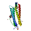
| ||||||||||||
|---|---|---|---|---|---|---|---|---|---|---|---|---|---|
| 1 | x 24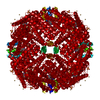
| ||||||||||||
| Unit cell |
| ||||||||||||
| Components on special symmetry positions |
|
-Components
| #1: Protein | / Ferritin L subunit Mass: 19872.428 Da / Num. of mol.: 1 / Source method: isolated from a natural source / Source: (natural) Equus caballus (horse) / Organ: Spleen / References: UniProt: P02791 | ||||||
|---|---|---|---|---|---|---|---|
| #2: Chemical | ChemComp-CD /   Mass: 112.411 Da / Num. of mol.: 6 / Source method: obtained synthetically / Formula: Cd Mass: 112.411 Da / Num. of mol.: 6 / Source method: obtained synthetically / Formula: Cd#3: Chemical | Sulfate  Mass: 96.063 Da / Num. of mol.: 2 / Source method: obtained synthetically / Formula: SO4 Mass: 96.063 Da / Num. of mol.: 2 / Source method: obtained synthetically / Formula: SO4#4: Chemical | ChemComp-RAV / | Pentobarbital  Mass: 226.272 Da / Num. of mol.: 1 / Source method: obtained synthetically / Formula: C11H18N2O3 Mass: 226.272 Da / Num. of mol.: 1 / Source method: obtained synthetically / Formula: C11H18N2O3#5: Water | ChemComp-HOH / | Water Mass: 18.015 Da / Num. of mol.: 196 / Source method: isolated from a natural source / Formula: H2O Mass: 18.015 Da / Num. of mol.: 196 / Source method: isolated from a natural source / Formula: H2O |
-Experimental details
-Experiment
| Experiment | Method: X-RAY DIFFRACTION / Number of used crystals: 1 |
|---|
- Sample preparation
| Crystal | Density Matthews: 3.3 Å3/Da / Density % sol: 62.3 % |
|---|---|
| Crystal grow | Temperature: 291 K / Method: vapor diffusion, hanging drop / pH: 7 Details: 0.2-1.6 M (NH4)2SO4 and 0.1-0.275 M CdSO4, 0.5 mM thiopental were mixed with equal volumes of apoferritin solution and equilibrated over 0.7-1 mL, VAPOR DIFFUSION, HANGING DROP, temperature 291K |
-Data collection
| Diffraction | Mean temperature: 110 K |
|---|---|
| Diffraction source | Source: SYNCHROTRON / Site: NSLS  / Beamline: X6A / Wavelength: 0.9774 Å / Beamline: X6A / Wavelength: 0.9774 Å |
| Radiation | Protocol: SINGLE WAVELENGTH / Monochromatic (M) / Laue (L): M / Scattering type: x-ray |
| Radiation wavelength | Wavelength: 0.9774 Å / Relative weight: 1 |
| Reflection | Resolution: 1.9→45.62 Å / Num. all: 21095 / Num. obs: 21095 / % possible obs: 100 % / Observed criterion σ(F): 38.9 / Observed criterion σ(I): 7.8 / Redundancy: 34.09 % / Biso Wilson estimate: 21.63 Å2 / Rmerge(I) obs: 0.109 / Net I/σ(I): 20 |
| Reflection shell | Resolution: 1.9→1.97 Å / Redundancy: 34.09 % / Rmerge(I) obs: 0.527 / Mean I/σ(I) obs: 7.8 / % possible all: 100 |
-Phasing
| Phasing | Method: molecular replacement | |||||||||
|---|---|---|---|---|---|---|---|---|---|---|
| Phasing MR | Rfactor: 56.24 / Model details: Phaser MODE: MR_AUTO
|
- Processing
| Software |
| |||||||||||||||||||||||||||||||||||||||||||||||||||||||||||||||||||||||||||||||||||||||||||||||||||||||||
|---|---|---|---|---|---|---|---|---|---|---|---|---|---|---|---|---|---|---|---|---|---|---|---|---|---|---|---|---|---|---|---|---|---|---|---|---|---|---|---|---|---|---|---|---|---|---|---|---|---|---|---|---|---|---|---|---|---|---|---|---|---|---|---|---|---|---|---|---|---|---|---|---|---|---|---|---|---|---|---|---|---|---|---|---|---|---|---|---|---|---|---|---|---|---|---|---|---|---|---|---|---|---|---|---|---|---|
| Refinement | Method to determine structure: molecular replacement, molecular replacement Resolution: 1.9→45.62 Å / Occupancy max: 1 / Occupancy min: 0.22 / SU ML: 0.2 / σ(F): 0.84 / Stereochemistry target values: ML
| |||||||||||||||||||||||||||||||||||||||||||||||||||||||||||||||||||||||||||||||||||||||||||||||||||||||||
| Solvent computation | Shrinkage radii: 1.06 Å / VDW probe radii: 1.3 Å / Solvent model: FLAT BULK SOLVENT MODEL / Bsol: 42.874 Å2 / ksol: 0.4 e/Å3 | |||||||||||||||||||||||||||||||||||||||||||||||||||||||||||||||||||||||||||||||||||||||||||||||||||||||||
| Displacement parameters | Biso max: 75.75 Å2 / Biso mean: 22.472 Å2 / Biso min: 11.86 Å2
| |||||||||||||||||||||||||||||||||||||||||||||||||||||||||||||||||||||||||||||||||||||||||||||||||||||||||
| Refinement step | Cycle: LAST / Resolution: 1.9→45.62 Å
| |||||||||||||||||||||||||||||||||||||||||||||||||||||||||||||||||||||||||||||||||||||||||||||||||||||||||
| Refine LS restraints |
| |||||||||||||||||||||||||||||||||||||||||||||||||||||||||||||||||||||||||||||||||||||||||||||||||||||||||
| LS refinement shell | Refine-ID: X-RAY DIFFRACTION / Total num. of bins used: 14
|
