 Movie
Movie Controller


+ Open data

- Basic information
| Entry | Database: PDB / ID: 3rab | ||||||
|---|---|---|---|---|---|---|---|
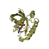
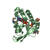
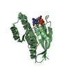
| Title | GPPNHP-BOUND RAB3A AT 2.0 A RESOLUTION | ||||||
 Components Components | PROTEIN (RAB3A) | ||||||
 Keywords Keywords |  HYDROLASE / G PROTEIN / VESICULAR TRAFFICKING / GTP HYDROLYSIS / RAB PROTEIN / NEUROTRANSMITTER RELEASE HYDROLASE / G PROTEIN / VESICULAR TRAFFICKING / GTP HYDROLYSIS / RAB PROTEIN / NEUROTRANSMITTER RELEASE | ||||||
| Function / homology |  Function and homology information Function and homology informationGDP-dissociation inhibitor binding / evoked neurotransmitter secretion / acrosomal vesicle exocytosis / regulation of presynaptic dense core granule exocytosis / RAB GEFs exchange GTP for GDP on RABs / positive regulation of regulated secretory pathway / regulation of plasma membrane repair / maintenance of presynaptic active zone structure / regulation of synaptic vesicle fusion to presynaptic active zone membrane / sensory perception of touch ...GDP-dissociation inhibitor binding / evoked neurotransmitter secretion / acrosomal vesicle exocytosis / regulation of presynaptic dense core granule exocytosis / RAB GEFs exchange GTP for GDP on RABs / positive regulation of regulated secretory pathway / regulation of plasma membrane repair / maintenance of presynaptic active zone structure / regulation of synaptic vesicle fusion to presynaptic active zone membrane / sensory perception of touch / RAB geranylgeranylation / Rab protein signal transduction / synaptic vesicle recycling / regulation of synaptic vesicle priming / Glutamate Neurotransmitter Release Cycle / Norepinephrine Neurotransmitter Release Cycle / Acetylcholine Neurotransmitter Release Cycle / Serotonin Neurotransmitter Release Cycle / GABA synthesis, release, reuptake and degradation / regulated exocytosis / Dopamine Neurotransmitter Release Cycle / synaptic vesicle clustering / neuromuscular synaptic transmission / synaptic vesicle maturation / GTP-dependent protein binding / respiratory system process / calcium-ion regulated exocytosis / myosin V binding / plasma membrane repair / lysosome localization / vesicle docking involved in exocytosis / regulation of exocytosis / regulation of short-term neuronal synaptic plasticity / insulin secretion / Neutrophil degranulation / synaptic vesicle transport / regulation of synaptic vesicle exocytosis / regulation of dopamine secretion / presynaptic active zone / exocytosis / synaptic vesicle exocytosis / ATPase activator activity / protein secretion / response to electrical stimulus / mitochondrion organization / axonogenesis / post-embryonic development / acrosomal vesicle / secretory granule / protein localization to plasma membrane / establishment of localization in cell / lung development / terminal bouton / synaptic vesicle membrane / synaptic vesicle / presynapse / protein-macromolecule adaptor activity / ATPase binding / postsynapse / lysosome / endosome / axon / GTPase activity / GTP binding / perinuclear region of cytoplasm / protein-containing complex / plasma membrane / cytosolSimilarity search - Function | ||||||
| Biological species |  Rattus norvegicus (Norway rat) Rattus norvegicus (Norway rat) | ||||||
| Method | X-RAY DIFFRACTION / MOLECULAR REPLACEMENT / Resolution: 2 Å | ||||||
 Authors Authors | Dumas, J.J. / Zhu, Z. / Connolly, J.L. / Lambright, D.G. | ||||||
 Citation Citation | Journal: Structure Fold.Des. / Year: 1999 Title: Structural basis of activation and GTP hydrolysis in Rab proteins. Authors: Dumas, J.J. / Zhu, Z. / Connolly, J.L. / Lambright, D.G. | ||||||
| History |
|
- Structure visualization
| Structure viewer | Molecule: MolmilJmol/JSmol |
|---|
- Downloads & links
-Download
| PDBx/mmCIF format | 3rab.cif.gz | 52.3 KB | Display | PDBx/mmCIF format |
|---|---|---|---|---|
| PDB format | pdb3rab.ent.gz | 34.6 KB | Display | PDB format |
| PDBx/mmJSON format | 3rab.json.gz | Tree view | PDBx/mmJSON format | |
| Others |  Other downloads Other downloads |
-Validation report
| Arichive directory | https://data.pdbj.org/pub/pdb/validation_reports/ra/3rabftp://data.pdbj.org/pub/pdb/validation_reports/ra/3rab | HTTPS FTP |
|---|
-Related structure data
| Related structure data | 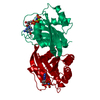 5p21S S: Starting model for refinement |
|---|---|
| Similar structure data |


-Links
 PDBj
PDBj


- Assembly
| Deposited unit | 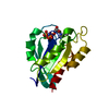
| ||||||||
|---|---|---|---|---|---|---|---|---|---|
| 1 |
| ||||||||
| Unit cell |
|
-Components
| #1: Protein | Mass: 19655.135 Da / Num. of mol.: 1 / Fragment: GTPASE DOMAIN Source method: isolated from a genetically manipulated source Source: (gene. exp.) Rattus norvegicus (Norway rat) / Organ: BRAIN / Plasmid: PGEX / Species (production host): Escherichia coli / Production host:  Escherichia coli BL21(DE3) (bacteria) / Strain (production host): BL21/DE3 / References: UniProt: P63012 Escherichia coli BL21(DE3) (bacteria) / Strain (production host): BL21/DE3 / References: UniProt: P63012 |
|---|---|
| #2: Chemical | ChemComp-MG /   Mass: 24.305 Da / Num. of mol.: 1 / Source method: obtained synthetically / Formula: Mg Mass: 24.305 Da / Num. of mol.: 1 / Source method: obtained synthetically / Formula: Mg |
| #3: Chemical | ChemComp-GNP / 5'-Guanylyl imidodiphosphate  Mass: 522.196 Da / Num. of mol.: 1 / Source method: obtained synthetically / Formula: C10H17N6O13P3 Mass: 522.196 Da / Num. of mol.: 1 / Source method: obtained synthetically / Formula: C10H17N6O13P3Comment: GppNHp, GMPPNP, energy-carrying molecule analogue*YM |
| #4: Water | ChemComp-HOH / Water Mass: 18.015 Da / Num. of mol.: 123 / Source method: isolated from a natural source / Formula: H2O Mass: 18.015 Da / Num. of mol.: 123 / Source method: isolated from a natural source / Formula: H2O |
-Experimental details
-Experiment
| Experiment | Method: X-RAY DIFFRACTION / Number of used crystals: 1 |
|---|
- Sample preparation
| Crystal | Density Matthews: 2.22 Å3/Da / Density % sol: 44 % | ||||||||||||||||||||||||||||||||||||||||||
|---|---|---|---|---|---|---|---|---|---|---|---|---|---|---|---|---|---|---|---|---|---|---|---|---|---|---|---|---|---|---|---|---|---|---|---|---|---|---|---|---|---|---|---|
| Crystal grow | pH: 6.5 Details: CRYSTALLIZATION CONDITIONS: 14%PEG-8000,50 MM NAMES, PH 6.5, 200 MM NACL | ||||||||||||||||||||||||||||||||||||||||||
| Crystal grow | *PLUS Temperature: 4 ℃ / Method: vapor diffusion, hanging dropDetails: drop consists of equal volume of protein and reservoir solutions | ||||||||||||||||||||||||||||||||||||||||||
| Components of the solutions | *PLUS
|
-Data collection
| Diffraction | Mean temperature: 100 K |
|---|---|
| Diffraction source | Source: ROTATING ANODE / Type: RIGAKU RU200 / Wavelength: 1.5418 |
| Detector | Type: MARRESEARCH / Detector: IMAGE PLATE / Date: Dec 1, 1997 / Details: MIRRORS |
| Radiation | Protocol: SINGLE WAVELENGTH / Monochromatic (M) / Laue (L): M / Scattering type: x-ray |
| Radiation wavelength | Wavelength: 1.5418 Å / Relative weight: 1 |
| Reflection | Resolution: 2→20 Å / Num. obs: 10595 / % possible obs: 99.5 % / Observed criterion σ(I): -3 / Rsym value: 0.063 / Net I/σ(I): 17.1 |
| Reflection shell | Resolution: 2→2.1 Å / Mean I/σ(I) obs: 6.3 / Rsym value: 0.197 / % possible all: 99.5 |
| Reflection | *PLUS Rmerge(I) obs: 0.063 |
| Reflection shell | *PLUS % possible obs: 99.5 % / Rmerge(I) obs: 0.197 |
- Processing
| Software |
| ||||||||||||||||||||||||||||||||||||||||||||||||||||||||||||
|---|---|---|---|---|---|---|---|---|---|---|---|---|---|---|---|---|---|---|---|---|---|---|---|---|---|---|---|---|---|---|---|---|---|---|---|---|---|---|---|---|---|---|---|---|---|---|---|---|---|---|---|---|---|---|---|---|---|---|---|---|---|
| Refinement | Method to determine structure: MOLECULAR REPLACEMENT Starting model: 5P21 Resolution: 2→8 Å / Data cutoff high absF: 0 / Data cutoff low absF: 0 / Cross valid method: THROUGHOUT / σ(F): 2
| ||||||||||||||||||||||||||||||||||||||||||||||||||||||||||||
| Displacement parameters | Biso mean: 13 Å2 | ||||||||||||||||||||||||||||||||||||||||||||||||||||||||||||
| Refinement step | Cycle: LAST / Resolution: 2→8 Å
| ||||||||||||||||||||||||||||||||||||||||||||||||||||||||||||
| Refine LS restraints |
| ||||||||||||||||||||||||||||||||||||||||||||||||||||||||||||
| LS refinement shell | Highest resolution: 2 Å /
| ||||||||||||||||||||||||||||||||||||||||||||||||||||||||||||
| Software | *PLUS Name: X-PLOR / Version: 3.1 / Classification: refinement | ||||||||||||||||||||||||||||||||||||||||||||||||||||||||||||
| Refinement | *PLUS Rfactor obs: 0.191 | ||||||||||||||||||||||||||||||||||||||||||||||||||||||||||||
| Solvent computation | *PLUS | ||||||||||||||||||||||||||||||||||||||||||||||||||||||||||||
| Displacement parameters | *PLUS | ||||||||||||||||||||||||||||||||||||||||||||||||||||||||||||
| Refine LS restraints | *PLUS
| ||||||||||||||||||||||||||||||||||||||||||||||||||||||||||||
| LS refinement shell | *PLUS Rfactor obs: 0.184 |
