 Movie
Movie Controller

[English] 日本語
 Yorodumi
- PDB-3r7g: Crystal structure of Spire KIND domain in complex with the tail o... -
+ Open data

- Basic information
| Entry | Database: PDB / ID: 3r7g | ||||||
|---|---|---|---|---|---|---|---|
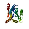
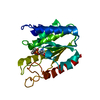
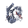
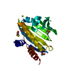
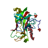
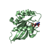
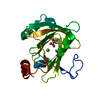
| Title | Crystal structure of Spire KIND domain in complex with the tail of FMN2 | ||||||
 Components Components |
| ||||||
 Keywords Keywords |  PROTEIN BINDING / C-lobe of protein kinases / actin nucleator / Fmn-family formins PROTEIN BINDING / C-lobe of protein kinases / actin nucleator / Fmn-family formins | ||||||
| Function / homology |  Function and homology information Function and homology informationhomologous chromosome movement towards spindle pole in meiosis I anaphase / formin-nucleated actin cable assembly / establishment of meiotic spindle localization / polar body extrusion after meiotic divisions / actin filament network formation / actin nucleation / Golgi vesicle transport / cleavage furrow formation / positive regulation of double-strand break repair / oogenesis ...homologous chromosome movement towards spindle pole in meiosis I anaphase / formin-nucleated actin cable assembly / establishment of meiotic spindle localization / polar body extrusion after meiotic divisions / actin filament network formation / actin nucleation / Golgi vesicle transport / cleavage furrow formation / positive regulation of double-strand break repair / oogenesis / positive regulation of mitochondrial fission / microvillus / intracellular transport / vesicle-mediated transport / actin filament polymerization / cytoplasmic vesicle membrane / negative regulation of protein catabolic process / spindle / cell migration / protein transport / actin binding / cell cortex / cellular response to hypoxia / actin cytoskeleton organization / mitochondrial outer membrane / cytoskeleton / intracellular signal transduction / innate immune response / DNA damage response / endoplasmic reticulum membrane / nucleolus / negative regulation of apoptotic process / perinuclear region of cytoplasm / endoplasmic reticulum / nucleoplasm / nucleus / plasma membrane / cytosol / cytoplasmSimilarity search - Function | ||||||
| Biological species |  Homo sapiens (human) Homo sapiens (human) | ||||||
| Method | X-RAY DIFFRACTION / MIR / Resolution: 2.2 Å | ||||||
 Authors Authors | Kreutz, B. / Vizcarra, C.L. / Rodal, A.A. / Toms, A.V. / Lu, J. / Quinlan, M.E. / Eck, M.J. | ||||||
 Citation Citation | Journal: Proc.Natl.Acad.Sci.USA / Year: 2011 Title: Structure of the Spire KIND domain and insights into its interaction with Fmn-family formins Authors: Vizcarra, C. / Kreutz, B. / Rodal, A. / Toms, A. / Lu, J. / Zheng, W. / Quinlan, M. / Eck, M. | ||||||
| History |
|
- Structure visualization
| Structure viewer | Molecule: MolmilJmol/JSmol |
|---|
- Downloads & links
-Download
| PDBx/mmCIF format | 3r7g.cif.gz | 50 KB | Display | PDBx/mmCIF format |
|---|---|---|---|---|
| PDB format | pdb3r7g.ent.gz | 35 KB | Display | PDB format |
| PDBx/mmJSON format | 3r7g.json.gz | Tree view | PDBx/mmJSON format | |
| Others |  Other downloads Other downloads |
-Validation report
| Arichive directory | https://data.pdbj.org/pub/pdb/validation_reports/r7/3r7gftp://data.pdbj.org/pub/pdb/validation_reports/r7/3r7g | HTTPS FTP |
|---|
-Related structure data


-Links
 PDBj
PDBj
- Assembly
| Deposited unit | 
| ||||||||
|---|---|---|---|---|---|---|---|---|---|
| 1 |
| ||||||||
| Unit cell |
|
-Components
| #1: Protein | Mass: 24027.785 Da / Num. of mol.: 1 / Fragment: KIND domain (UNP residues 20-237) Source method: isolated from a genetically manipulated source Details: Spir1 KIND 20-237 / Source: (gene. exp.) Homo sapiens (human) / Gene: SPIRE1, KIAA1135, SPIR1 / Plasmid: pET HisTT / Production host:  Escherichia coli (E. coli) / Strain (production host): BL21DE3 / References: UniProt: Q08AE8 Escherichia coli (E. coli) / Strain (production host): BL21DE3 / References: UniProt: Q08AE8 |
|---|---|
| #2: Protein/peptide | Formins Mass: 2538.105 Da / Num. of mol.: 1 / Fragment: FSI domain (UNP residues 1701-1722) Source method: isolated from a genetically manipulated source Details: Fmn2 1700-1722 / Source: (gene. exp.) Homo sapiens (human) / Gene: FMN2 / Plasmid: pET GST / Production host: Escherichia coli (E. coli) / Strain (production host): BL21DE3 / References: UniProt: Q9NZ56 |
| #3: Water | ChemComp-HOH / Water Mass: 18.015 Da / Num. of mol.: 85 / Source method: isolated from a natural source / Formula: H2O Mass: 18.015 Da / Num. of mol.: 85 / Source method: isolated from a natural source / Formula: H2O |
-Experimental details
-Experiment
| Experiment | Method: X-RAY DIFFRACTION |
|---|
- Sample preparation
| Crystal | Density Matthews: 2.42 Å3/Da / Density % sol: 49.13 % |
|---|---|
| Crystal grow | Temperature: 277 K / Method: vapor diffusion / pH: 6 / Details: PEG400, pH 6.0, VAPOR DIFFUSION, temperature 277K |
-Data collection
| Diffraction | Mean temperature: 295 K |
|---|---|
| Diffraction source | Source: ROTATING ANODE / Type: RIGAKU RU300 / Wavelength: 1.54 Å |
| Detector | Type: MAR scanner 345 mm plate / Detector: IMAGE PLATE / Date: Sep 8, 2008 |
| Radiation | Protocol: SINGLE WAVELENGTH / Monochromatic (M) / Laue (L): M / Scattering type: x-ray |
| Radiation wavelength | Wavelength: 1.54 Å / Relative weight: 1 |
| Reflection | Resolution: 2.2→45 Å / Num. obs: 12200 / % possible obs: 95.5 % / Rmerge(I) obs: 0.081 |
- Processing
| Software |
| ||||||||||||||||||||||||||||||||||||||||||||||||||||||||||||||||||||||||||||||||||||||||||||||||||||||||||||||||||||||||||||||||||||||||||||||||||||||||||||||||||||||||||
|---|---|---|---|---|---|---|---|---|---|---|---|---|---|---|---|---|---|---|---|---|---|---|---|---|---|---|---|---|---|---|---|---|---|---|---|---|---|---|---|---|---|---|---|---|---|---|---|---|---|---|---|---|---|---|---|---|---|---|---|---|---|---|---|---|---|---|---|---|---|---|---|---|---|---|---|---|---|---|---|---|---|---|---|---|---|---|---|---|---|---|---|---|---|---|---|---|---|---|---|---|---|---|---|---|---|---|---|---|---|---|---|---|---|---|---|---|---|---|---|---|---|---|---|---|---|---|---|---|---|---|---|---|---|---|---|---|---|---|---|---|---|---|---|---|---|---|---|---|---|---|---|---|---|---|---|---|---|---|---|---|---|---|---|---|---|---|---|---|---|---|---|
| Refinement | Method to determine structure: MIR / Resolution: 2.2→38.8 Å / Cor.coef. Fo:Fc: 0.949 / Cor.coef. Fo:Fc free: 0.937 / SU B: 4.66 / SU ML: 0.118 / Cross valid method: THROUGHOUT / ESU R Free: 0.181 / Stereochemistry target values: MAXIMUM LIKELIHOOD
| ||||||||||||||||||||||||||||||||||||||||||||||||||||||||||||||||||||||||||||||||||||||||||||||||||||||||||||||||||||||||||||||||||||||||||||||||||||||||||||||||||||||||||
| Solvent computation | Ion probe radii: 0.8 Å / Shrinkage radii: 0.8 Å / VDW probe radii: 1.4 Å / Solvent model: MASK | ||||||||||||||||||||||||||||||||||||||||||||||||||||||||||||||||||||||||||||||||||||||||||||||||||||||||||||||||||||||||||||||||||||||||||||||||||||||||||||||||||||||||||
| Displacement parameters | Biso mean: 32.877 Å2
| ||||||||||||||||||||||||||||||||||||||||||||||||||||||||||||||||||||||||||||||||||||||||||||||||||||||||||||||||||||||||||||||||||||||||||||||||||||||||||||||||||||||||||
| Refinement step | Cycle: LAST / Resolution: 2.2→38.8 Å
| ||||||||||||||||||||||||||||||||||||||||||||||||||||||||||||||||||||||||||||||||||||||||||||||||||||||||||||||||||||||||||||||||||||||||||||||||||||||||||||||||||||||||||
| Refine LS restraints |
| ||||||||||||||||||||||||||||||||||||||||||||||||||||||||||||||||||||||||||||||||||||||||||||||||||||||||||||||||||||||||||||||||||||||||||||||||||||||||||||||||||||||||||
| LS refinement shell | Resolution: 2.202→2.259 Å / Total num. of bins used: 20
|
