 Movie
Movie Controller

[English] 日本語
 Yorodumi
- PDB-3r74: Crystal structure of 2-amino-2-desoxyisochorismate synthase (ADIC... -
+ Open data

- Basic information
| Entry | Database: PDB / ID: 3r74 | ||||||
|---|---|---|---|---|---|---|---|
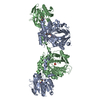
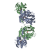
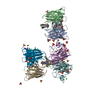
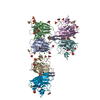
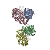
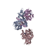
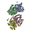
| Title | Crystal structure of 2-amino-2-desoxyisochorismate synthase (ADIC) synthase PhzE from Burkholderia lata 383 | ||||||
 Components Components | Anthranilate/para-aminobenzoate synthases component I | ||||||
 Keywords Keywords |  LYASE / BIOSYNTHETIC PROTEIN / ammonia channel / chorismate / type 1 glutamine amidotransferase / phenazine biosynthesis / SYNTHASE LYASE / BIOSYNTHETIC PROTEIN / ammonia channel / chorismate / type 1 glutamine amidotransferase / phenazine biosynthesis / SYNTHASE | ||||||
| Function / homology |  Function and homology informationanthranilate synthase / anthranilate synthase activity / tryptophan biosynthetic process / glutamine metabolic process / metal ion binding Function and homology informationanthranilate synthase / anthranilate synthase activity / tryptophan biosynthetic process / glutamine metabolic process / metal ion bindingSimilarity search - Function | ||||||
| Biological species |  Burkholderia sp. (bacteria) Burkholderia sp. (bacteria) | ||||||
| Method | X-RAY DIFFRACTION / SYNCHROTRON / MAD / Resolution: 2.9 Å | ||||||
 Authors Authors | Li, Q.A. / Mavrodi, D.V. / Thomashow, L.S. / Roessle, M. / Blankenfeldt, W. | ||||||
 Citation Citation | Journal: J.Biol.Chem. / Year: 2011 Title: Ligand Binding Induces an Ammonia Channel in 2-Amino-2-desoxyisochorismate (ADIC) Synthase PhzE. Authors: Li, Q.A. / Mavrodi, D.V. / Thomashow, L.S. / Roessle, M. / Blankenfeldt, W. | ||||||
| History |
|
- Structure visualization
| Structure viewer | Molecule: MolmilJmol/JSmol |
|---|
- Downloads & links
-Download
| PDBx/mmCIF format | 3r74.cif.gz | 467.3 KB | Display | PDBx/mmCIF format |
|---|---|---|---|---|
| PDB format | pdb3r74.ent.gz | 387.9 KB | Display | PDB format |
| PDBx/mmJSON format | 3r74.json.gz | Tree view | PDBx/mmJSON format | |
| Others |  Other downloads Other downloads |
-Validation report
| Arichive directory | https://data.pdbj.org/pub/pdb/validation_reports/r7/3r74ftp://data.pdbj.org/pub/pdb/validation_reports/r7/3r74 | HTTPS FTP |
|---|
-Related structure data


-Links
 PDBj
PDBj

- Assembly
| Deposited unit | 
| ||||||||||||
|---|---|---|---|---|---|---|---|---|---|---|---|---|---|
| 1 |
| ||||||||||||
| Unit cell |
| ||||||||||||
| Components on special symmetry positions |
|
-Components
| #1: Protein | Mass: 70163.234 Da / Num. of mol.: 2 Source method: isolated from a genetically manipulated source Source: (gene. exp.) Burkholderia sp. (bacteria) / Strain: 383 / Gene: Bcep18194_B1570 / Plasmid: pET19modTEV / Production host: Escherichia coli (E. coli) / Strain (production host): Rosetta 2 pLys S / References: UniProt: Q396C7, anthranilate synthase#2: Water | ChemComp-HOH / | Water Mass: 18.015 Da / Num. of mol.: 80 / Source method: isolated from a natural source / Formula: H2O Mass: 18.015 Da / Num. of mol.: 80 / Source method: isolated from a natural source / Formula: H2O |
|---|
-Experimental details
-Experiment
| Experiment | Method: X-RAY DIFFRACTION / Number of used crystals: 1 |
|---|
- Sample preparation
| Crystal | Density Matthews: 3.46 Å3/Da / Density % sol: 64.5 % |
|---|---|
| Crystal grow | Temperature: 298 K / Method: vapor diffusion, hanging drop / pH: 7 Details: 0.1 M Bis-TRIS propane, 0.2 M KSCN, 22% (w/v) PEG 3350, 1 mM Mg-chloride, 20 mM chorismate, pH 7.0, vapor diffusion, hanging drop, temperature 298K |
-Data collection
| Diffraction | Mean temperature: 100 K | ||||||||||||||||||||||||||||||||||||||||||||||||||||||||||||||||||||||||||||||||||||||||||||||||||
|---|---|---|---|---|---|---|---|---|---|---|---|---|---|---|---|---|---|---|---|---|---|---|---|---|---|---|---|---|---|---|---|---|---|---|---|---|---|---|---|---|---|---|---|---|---|---|---|---|---|---|---|---|---|---|---|---|---|---|---|---|---|---|---|---|---|---|---|---|---|---|---|---|---|---|---|---|---|---|---|---|---|---|---|---|---|---|---|---|---|---|---|---|---|---|---|---|---|---|---|
| Diffraction source | Source: SYNCHROTRON / Site: SLS  / Beamline: X10SA / Wavelength: 1.0, 0.97895, 0.97957, 0.97793 / Beamline: X10SA / Wavelength: 1.0, 0.97895, 0.97957, 0.97793 | ||||||||||||||||||||||||||||||||||||||||||||||||||||||||||||||||||||||||||||||||||||||||||||||||||
| Detector | Type: MARMOSAIC 225 mm CCD / Detector: CCD / Date: Nov 7, 2007 / Details: SI(111) | ||||||||||||||||||||||||||||||||||||||||||||||||||||||||||||||||||||||||||||||||||||||||||||||||||
| Radiation | Monochromator: SI(111) MONOCHROMATOR / Protocol: MAD / Monochromatic (M) / Laue (L): M / Scattering type: x-ray | ||||||||||||||||||||||||||||||||||||||||||||||||||||||||||||||||||||||||||||||||||||||||||||||||||
| Radiation wavelength |
| ||||||||||||||||||||||||||||||||||||||||||||||||||||||||||||||||||||||||||||||||||||||||||||||||||
| Reflection | Resolution: 2.9→19.924 Å / Num. obs: 42476 / % possible obs: 99.6 % / Observed criterion σ(I): -3 / Biso Wilson estimate: 60.355 Å2 / Rmerge(I) obs: 0.062 / Net I/σ(I): 20.07 | ||||||||||||||||||||||||||||||||||||||||||||||||||||||||||||||||||||||||||||||||||||||||||||||||||
| Reflection shell | Diffraction-ID: 1
|
-Phasing
| Phasing | Method: MAD |
|---|
- Processing
| Software |
| |||||||||||||||||||||||||||||||||||||||||||||||||||||||||||||||||||||||||||||||||||||||||||||||||||||||||||||||||||||||||||||
|---|---|---|---|---|---|---|---|---|---|---|---|---|---|---|---|---|---|---|---|---|---|---|---|---|---|---|---|---|---|---|---|---|---|---|---|---|---|---|---|---|---|---|---|---|---|---|---|---|---|---|---|---|---|---|---|---|---|---|---|---|---|---|---|---|---|---|---|---|---|---|---|---|---|---|---|---|---|---|---|---|---|---|---|---|---|---|---|---|---|---|---|---|---|---|---|---|---|---|---|---|---|---|---|---|---|---|---|---|---|---|---|---|---|---|---|---|---|---|---|---|---|---|---|---|---|---|
| Refinement | Method to determine structure: MAD / Resolution: 2.9→19.92 Å / Cor.coef. Fo:Fc: 0.949 / Cor.coef. Fo:Fc free: 0.922 / Occupancy max: 1 / Occupancy min: 0.5 / SU B: 26.439 / SU ML: 0.237 / Cross valid method: THROUGHOUT / σ(F): 0 / ESU R: 0.885 / ESU R Free: 0.326 / Stereochemistry target values: MAXIMUM LIKELIHOOD / Details: HYDROGENS HAVE BEEN USED IF PRESENT IN THE INPUT
| |||||||||||||||||||||||||||||||||||||||||||||||||||||||||||||||||||||||||||||||||||||||||||||||||||||||||||||||||||||||||||||
| Solvent computation | Ion probe radii: 0.8 Å / Shrinkage radii: 0.8 Å / VDW probe radii: 1.2 Å / Solvent model: BABINET MODEL WITH MASK | |||||||||||||||||||||||||||||||||||||||||||||||||||||||||||||||||||||||||||||||||||||||||||||||||||||||||||||||||||||||||||||
| Displacement parameters | Biso max: 206.44 Å2 / Biso mean: 76.0686 Å2 / Biso min: 23.4 Å2 | |||||||||||||||||||||||||||||||||||||||||||||||||||||||||||||||||||||||||||||||||||||||||||||||||||||||||||||||||||||||||||||
| Refinement step | Cycle: LAST / Resolution: 2.9→19.92 Å
| |||||||||||||||||||||||||||||||||||||||||||||||||||||||||||||||||||||||||||||||||||||||||||||||||||||||||||||||||||||||||||||
| Refine LS restraints |
| |||||||||||||||||||||||||||||||||||||||||||||||||||||||||||||||||||||||||||||||||||||||||||||||||||||||||||||||||||||||||||||
| LS refinement shell | Resolution: 2.9→2.974 Å / Total num. of bins used: 20
| |||||||||||||||||||||||||||||||||||||||||||||||||||||||||||||||||||||||||||||||||||||||||||||||||||||||||||||||||||||||||||||
| Refinement TLS params. | Method: refined / Refine-ID: X-RAY DIFFRACTION
| |||||||||||||||||||||||||||||||||||||||||||||||||||||||||||||||||||||||||||||||||||||||||||||||||||||||||||||||||||||||||||||
| Refinement TLS group |
|
