 Movie
Movie Controller


+ Open data

- Basic information
| Entry | Database: PDB / ID: 3r6p | ||||||
|---|---|---|---|---|---|---|---|
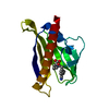
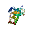
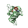
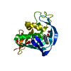
| Title | Crystal structure of abscisic acid-bound PYL10 | ||||||
 Components Components | Abscisic acid receptor PYL10 | ||||||
 Keywords Keywords |  HORMONE RECEPTOR / Arabidopsis Proteins Receptors / Abscisic Acid binding HORMONE RECEPTOR / Arabidopsis Proteins Receptors / Abscisic Acid binding | ||||||
| Function / homology |  Function and homology informationregulation of protein serine/threonine phosphatase activity / abscisic acid binding / abscisic acid-activated signaling pathway / protein phosphatase inhibitor activity / signaling receptor activity / protein homodimerization activity / nucleus / plasma membrane / cytoplasm Function and homology informationregulation of protein serine/threonine phosphatase activity / abscisic acid binding / abscisic acid-activated signaling pathway / protein phosphatase inhibitor activity / signaling receptor activity / protein homodimerization activity / nucleus / plasma membrane / cytoplasmSimilarity search - Function | ||||||
| Biological species |  Arabidopsis thaliana (thale cress) Arabidopsis thaliana (thale cress) | ||||||
| Method | X-RAY DIFFRACTION / SYNCHROTRON / MOLECULAR REPLACEMENT / Resolution: 2.7 Å | ||||||
 Authors Authors | Sun, D.M. / Wu, M.H. / Wang, H.P. / Zang, J.Y. / Tian, C.L. | ||||||
 Citation Citation | Journal: To be Published Title: Crystal structure of abscisic acid-bound PYL10 Authors: Sun, D.M. / Wu, M.H. / Wang, H.P. / Zang, J.Y. / Tian, C.L. | ||||||
| History |
|
- Structure visualization
| Structure viewer | Molecule: MolmilJmol/JSmol |
|---|
- Downloads & links
-Download
| PDBx/mmCIF format | 3r6p.cif.gz | 44.6 KB | Display | PDBx/mmCIF format |
|---|---|---|---|---|
| PDB format | pdb3r6p.ent.gz | 30.2 KB | Display | PDB format |
| PDBx/mmJSON format | 3r6p.json.gz | Tree view | PDBx/mmJSON format | |
| Others |  Other downloads Other downloads |
-Validation report
| Arichive directory | https://data.pdbj.org/pub/pdb/validation_reports/r6/3r6pftp://data.pdbj.org/pub/pdb/validation_reports/r6/3r6p | HTTPS FTP |
|---|
-Related structure data
| Related structure data |  3uqhS S: Starting model for refinement |
|---|---|
| Similar structure data |


-Links
 PDBj
PDBj- Assembly
| Deposited unit | 
| ||||||||
|---|---|---|---|---|---|---|---|---|---|
| 1 |
| ||||||||
| 2 | 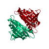
| ||||||||
| Unit cell |
|
-Components
| #1: Protein | Mass: 21745.762 Da / Num. of mol.: 1 Source method: isolated from a genetically manipulated source Source: (gene. exp.) Arabidopsis thaliana (thale cress) / Gene: PYL10 / Plasmid: pET21b / Production host:  Escherichia coli (E. coli) / Strain (production host): BL21(DE3) / References: UniProt: Q8H1R0 Escherichia coli (E. coli) / Strain (production host): BL21(DE3) / References: UniProt: Q8H1R0 |
|---|---|
| #2: Chemical | ChemComp-A8S / (Abscisic acid  Mass: 264.317 Da / Num. of mol.: 1 / Source method: obtained synthetically / Formula: C15H20O4 / Comment: hormone*YM Mass: 264.317 Da / Num. of mol.: 1 / Source method: obtained synthetically / Formula: C15H20O4 / Comment: hormone*YM |
| #3: Water | ChemComp-HOH / Water Mass: 18.015 Da / Num. of mol.: 11 / Source method: isolated from a natural source / Formula: H2O Mass: 18.015 Da / Num. of mol.: 11 / Source method: isolated from a natural source / Formula: H2O |
-Experimental details
-Experiment
| Experiment | Method: X-RAY DIFFRACTION / Number of used crystals: 1 |
|---|
- Sample preparation
| Crystal | Density Matthews: 1.96 Å3/Da / Density % sol: 37.39 % |
|---|---|
| Crystal grow | Temperature: 295 K / Method: vapor diffusion, sitting drop / pH: 6.5 Details: 20% PEG monoethyl ether 5000, 0.1M Bis-Tris, pH 6.5, VAPOR DIFFUSION, SITTING DROP, temperature 295K |
-Data collection
| Diffraction | Mean temperature: 100 K |
|---|---|
| Diffraction source | Source: SYNCHROTRON / Site: SSRF  / Beamline: BL17U / Beamline: BL17U |
| Detector | Date: Mar 16, 2011 |
| Radiation | Protocol: SINGLE WAVELENGTH / Monochromatic (M) / Laue (L): M / Scattering type: x-ray |
| Radiation wavelength | Relative weight: 1 |
| Reflection | Resolution: 2.7→50 Å / Num. obs: 4929 |
- Processing
| Software |
| |||||||||||||||||||||||||||||||||||||||||||||
|---|---|---|---|---|---|---|---|---|---|---|---|---|---|---|---|---|---|---|---|---|---|---|---|---|---|---|---|---|---|---|---|---|---|---|---|---|---|---|---|---|---|---|---|---|---|---|
| Refinement | Method to determine structure: MOLECULAR REPLACEMENT Starting model: 3UQH Resolution: 2.7→50 Å / Cor.coef. Fo:Fc: 0.933 / Cor.coef. Fo:Fc free: 0.924 / Occupancy max: 1 / Occupancy min: 0.05 / Cross valid method: THROUGHOUT / σ(F): 0 / ESU R Free: 0.411 / Stereochemistry target values: MAXIMUM LIKELIHOOD Details: HYDROGENS HAVE BEEN USED IF PRESENT IN THE INPUT U VALUES: REFINED INDIVIDUALLY
| |||||||||||||||||||||||||||||||||||||||||||||
| Solvent computation | Ion probe radii: 0.8 Å / Shrinkage radii: 0.8 Å / VDW probe radii: 1.2 Å / Solvent model: MASK | |||||||||||||||||||||||||||||||||||||||||||||
| Displacement parameters | Biso max: 109.56 Å2 / Biso mean: 67.6368 Å2 / Biso min: 47.73 Å2
| |||||||||||||||||||||||||||||||||||||||||||||
| Refinement step | Cycle: LAST / Resolution: 2.7→50 Å
| |||||||||||||||||||||||||||||||||||||||||||||
| Refine LS restraints |
| |||||||||||||||||||||||||||||||||||||||||||||
| LS refinement shell | Resolution: 2.697→2.767 Å / Total num. of bins used: 20
|
