 Movie
Movie Controller

[English] 日本語
 Yorodumi
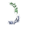
- PDB-3r2p: 2.2 Angstrom Crystal Structure of C Terminal Truncated Human Apol... -
+ Open data

- Basic information
| Entry | Database: PDB / ID: 3r2p | ||||||
|---|---|---|---|---|---|---|---|
| Title | 2.2 Angstrom Crystal Structure of C Terminal Truncated Human Apolipoprotein A-I Reveals the Assembly of HDL by Dimerization. | ||||||
 Components Components | Apolipoprotein A-I Apolipoprotein AI Apolipoprotein AI | ||||||
 Keywords Keywords | LIPID TRANSPORT / amphipathic alpha-helix / major protein of high density lipoprotein (HDL) / lipid binding / plasma | ||||||
| Function / homology |  Function and homology information Function and homology informationDefective ABCA1 causes TGD / high-density lipoprotein particle receptor binding / Scavenging by Class B Receptors / HDL clearance / spherical high-density lipoprotein particle / positive regulation of hydrolase activity / regulation of intestinal cholesterol absorption / negative regulation of response to cytokine stimulus / protein oxidation / vitamin transport ...Defective ABCA1 causes TGD / high-density lipoprotein particle receptor binding / Scavenging by Class B Receptors / HDL clearance / spherical high-density lipoprotein particle / positive regulation of hydrolase activity / regulation of intestinal cholesterol absorption / negative regulation of response to cytokine stimulus / protein oxidation / vitamin transport / high-density lipoprotein particle binding / cholesterol import / ABC transporters in lipid homeostasis / negative regulation of heterotypic cell-cell adhesion / blood vessel endothelial cell migration / apolipoprotein receptor binding / negative regulation of cell adhesion molecule production / negative regulation of cytokine production involved in immune response / apolipoprotein A-I receptor binding / HDL assembly / peptidyl-methionine modification / negative regulation of very-low-density lipoprotein particle remodeling / phosphatidylcholine biosynthetic process / glucocorticoid metabolic process / Chylomicron remodeling / phosphatidylcholine-sterol O-acyltransferase activator activity / phosphatidylcholine metabolic process / positive regulation of phospholipid efflux / lipid storage / phospholipid homeostasis / Chylomicron assembly / positive regulation of cholesterol metabolic process / high-density lipoprotein particle remodeling / high-density lipoprotein particle clearance / phospholipid efflux / cholesterol transfer activity / reverse cholesterol transport / cholesterol transport / chemorepellent activity / high-density lipoprotein particle assembly / very-low-density lipoprotein particle / positive regulation of CoA-transferase activity / lipoprotein biosynthetic process / high-density lipoprotein particle / endothelial cell proliferation / regulation of Cdc42 protein signal transduction / triglyceride homeostasis / HDL remodeling / cholesterol efflux / Scavenging by Class A Receptors / cholesterol binding / negative regulation of interleukin-1 beta production / positive regulation of Rho protein signal transduction / negative chemotaxis / adrenal gland development / cholesterol biosynthetic process / endocytic vesicle / positive regulation of cholesterol efflux / negative regulation of tumor necrosis factor-mediated signaling pathway / Scavenging of heme from plasma / positive regulation of phagocytosis / positive regulation of substrate adhesion-dependent cell spreading / Retinoid metabolism and transport / positive regulation of stress fiber assembly / endocytic vesicle lumen / heat shock protein binding / cholesterol metabolic process / cholesterol homeostasis / integrin-mediated signaling pathway / Post-translational protein phosphorylation / regulation of protein phosphorylation / phospholipid binding / Heme signaling / PPARA activates gene expression / negative regulation of inflammatory response / Regulation of Insulin-like Growth Factor (IGF) transport and uptake by Insulin-like Growth Factor Binding Proteins (IGFBPs) / extracellular vesicle / Platelet degranulation / amyloid-beta binding / cytoplasmic vesicle / collagen-containing extracellular matrix / secretory granule lumen / blood microparticle / protein stabilization / early endosome / G protein-coupled receptor signaling pathway / Amyloid fiber formation / endoplasmic reticulum lumen / signaling receptor binding / enzyme binding / protein homodimerization activity / extracellular space / extracellular exosome / extracellular region / identical protein binding / plasma membrane / cytosolSimilarity search - Function | ||||||
| Biological species |  Homo sapiens (human) Homo sapiens (human) | ||||||
| Method | X-RAY DIFFRACTION / SYNCHROTRON / MOLECULAR REPLACEMENT / Resolution: 2.2045 Å | ||||||
 Authors Authors | Mei, X. / Atkinson, D. | ||||||
 Citation Citation | Journal: J.Biol.Chem. / Year: 2011 Title: Crystal Structure of C-terminal Truncated Apolipoprotein A-I Reveals the Assembly of High Density Lipoprotein (HDL) by Dimerization. Authors: Mei, X. / Atkinson, D. | ||||||
| History |
|
- Structure visualization
| Structure viewer | Molecule: MolmilJmol/JSmol |
|---|
- Downloads & links
-Download
| PDBx/mmCIF format | 3r2p.cif.gz | 87.1 KB | Display | PDBx/mmCIF format |
|---|---|---|---|---|
| PDB format | pdb3r2p.ent.gz | 67.6 KB | Display | PDB format |
| PDBx/mmJSON format | 3r2p.json.gz | Tree view | PDBx/mmJSON format | |
| Others |  Other downloads Other downloads |
-Validation report
| Arichive directory | https://data.pdbj.org/pub/pdb/validation_reports/r2/3r2pftp://data.pdbj.org/pub/pdb/validation_reports/r2/3r2p | HTTPS FTP |
|---|
-Related structure data
| Similar structure data |
|---|
-Links
 PDBj
PDBj


- Assembly
| Deposited unit | 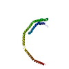
| ||||||||
|---|---|---|---|---|---|---|---|---|---|
| 1 | 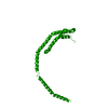
| ||||||||
| Unit cell |
| ||||||||
| Details | The biological assembly is a dimer generated from the monomer in the asymmetric unit by the operations: -x, -y, z and x, y+1, Z |
-Components
| #1: Protein | Apolipoprotein AI / Apo-AI / ApoA-I / Apolipoprotein A1 / Apolipoprotein A-I(1-242) Mass: 21656.234 Da / Num. of mol.: 1 / Fragment: N-terminal domain (UNP 25-208) Source method: isolated from a genetically manipulated source Source: (gene. exp.) Homo sapiens (human) / Gene: APOA1 / Plasmid: pDEST-His6-MBP / Production host:  Escherichia coli (E. coli) / Strain (production host): BL21(DE3) CodonPlus-RIL / References: UniProt: P02647 Escherichia coli (E. coli) / Strain (production host): BL21(DE3) CodonPlus-RIL / References: UniProt: P02647 |
|---|---|
| #2: Water | ChemComp-HOH / Water Mass: 18.015 Da / Num. of mol.: 38 / Source method: isolated from a natural source / Formula: H2O Mass: 18.015 Da / Num. of mol.: 38 / Source method: isolated from a natural source / Formula: H2O |
-Experimental details
-Experiment
| Experiment | Method: X-RAY DIFFRACTION / Number of used crystals: 1 |
|---|
- Sample preparation
| Crystal | Density Matthews: 2.63 Å3/Da / Density % sol: 53.16 % |
|---|---|
| Crystal grow | Temperature: 298 K / Method: vapor diffusion, hanging drop / pH: 6.6 Details: 0.15M potassium bromide, 30% PEG 2000 MME, pH 6.6, VAPOR DIFFUSION, HANGING DROP, temperature 298K |
-Data collection
| Diffraction | Mean temperature: 100 K |
|---|---|
| Diffraction source | Source: SYNCHROTRON / Site: NSLS  / Beamline: X4C / Wavelength: 0.91994 Å / Beamline: X4C / Wavelength: 0.91994 Å |
| Detector | Type: MAR CCD 165 mm / Detector: CCD / Date: Apr 15, 2010 |
| Radiation | Monochromator: Si 111 channel / Protocol: SINGLE WAVELENGTH / Monochromatic (M) / Laue (L): M / Scattering type: x-ray |
| Radiation wavelength | Wavelength: 0.91994 Å / Relative weight: 1 |
| Reflection | Resolution: 2.2→50 Å / Num. all: 11930 / Num. obs: 11930 / % possible obs: 98.2 % / Observed criterion σ(F): 2.95 / Observed criterion σ(I): 2.95 / Redundancy: 6.4 % / Biso Wilson estimate: 37.46 Å2 / Rmerge(I) obs: 0.074 / Net I/σ(I): 21.017 |
| Reflection shell | Resolution: 2.2→2.24 Å / Redundancy: 4.2 % / Rmerge(I) obs: 0.395 / Mean I/σ(I) obs: 2.95 / Num. unique all: 532 / % possible all: 89.4 |
- Processing
| Software |
| |||||||||||||||||||||||||||||||||||||||||||||||||||||||||||||||||||||||||||||||||||||||||||||||||||||||||||||||||||||||||||||
|---|---|---|---|---|---|---|---|---|---|---|---|---|---|---|---|---|---|---|---|---|---|---|---|---|---|---|---|---|---|---|---|---|---|---|---|---|---|---|---|---|---|---|---|---|---|---|---|---|---|---|---|---|---|---|---|---|---|---|---|---|---|---|---|---|---|---|---|---|---|---|---|---|---|---|---|---|---|---|---|---|---|---|---|---|---|---|---|---|---|---|---|---|---|---|---|---|---|---|---|---|---|---|---|---|---|---|---|---|---|---|---|---|---|---|---|---|---|---|---|---|---|---|---|---|---|---|
| Refinement | Method to determine structure: MOLECULAR REPLACEMENT Starting model: Use the Se-Met derivative to obtain the starting model from SAD experiment. Resolution: 2.2045→34.788 Å / SU ML: 0.33 / Isotropic thermal model: Mixed individual ADP with TLS / σ(F): 0 / Phase error: 33.52 / Stereochemistry target values: ML
| |||||||||||||||||||||||||||||||||||||||||||||||||||||||||||||||||||||||||||||||||||||||||||||||||||||||||||||||||||||||||||||
| Solvent computation | Shrinkage radii: 0.83 Å / VDW probe radii: 1.1 Å / Solvent model: FLAT BULK SOLVENT MODEL / Bsol: 47.738 Å2 / ksol: 0.332 e/Å3 | |||||||||||||||||||||||||||||||||||||||||||||||||||||||||||||||||||||||||||||||||||||||||||||||||||||||||||||||||||||||||||||
| Displacement parameters | Biso mean: 63.7952 Å2
| |||||||||||||||||||||||||||||||||||||||||||||||||||||||||||||||||||||||||||||||||||||||||||||||||||||||||||||||||||||||||||||
| Refinement step | Cycle: LAST / Resolution: 2.2045→34.788 Å
| |||||||||||||||||||||||||||||||||||||||||||||||||||||||||||||||||||||||||||||||||||||||||||||||||||||||||||||||||||||||||||||
| Refine LS restraints |
| |||||||||||||||||||||||||||||||||||||||||||||||||||||||||||||||||||||||||||||||||||||||||||||||||||||||||||||||||||||||||||||
| LS refinement shell | Refine-ID: X-RAY DIFFRACTION
| |||||||||||||||||||||||||||||||||||||||||||||||||||||||||||||||||||||||||||||||||||||||||||||||||||||||||||||||||||||||||||||
| Refinement TLS params. | Method: refined / Refine-ID: X-RAY DIFFRACTION
| |||||||||||||||||||||||||||||||||||||||||||||||||||||||||||||||||||||||||||||||||||||||||||||||||||||||||||||||||||||||||||||
| Refinement TLS group |
|
