 Movie
Movie Controller

[English] 日本語
 Yorodumi
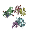
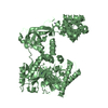
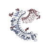
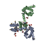
- PDB-3r0q: A Uniquely Open Conformation Revealed in the Crystal Structure of... -
+ Open data

- Basic information
| Entry | Database: PDB / ID: 3r0q | ||||||
|---|---|---|---|---|---|---|---|
| Title | A Uniquely Open Conformation Revealed in the Crystal Structure of Arabidopsis Thaliana Protein Arginine Methyltransferase 10 | ||||||
 Components Components | Probable protein arginine N-methyltransferase 4.2 Probability Probability | ||||||
 Keywords Keywords | TRANSFERASE / Arginine methyltransferase / Methylation | ||||||
| Function / homology |  Function and homology information: / peptidyl-arginine methylation, to asymmetrical-dimethyl arginine / vegetative to reproductive phase transition of meristem / protein-arginine omega-N monomethyltransferase activity / type I protein arginine methyltransferase / protein-arginine omega-N asymmetric methyltransferase activity / histone arginine N-methyltransferase activity / cytosol Function and homology information: / peptidyl-arginine methylation, to asymmetrical-dimethyl arginine / vegetative to reproductive phase transition of meristem / protein-arginine omega-N monomethyltransferase activity / type I protein arginine methyltransferase / protein-arginine omega-N asymmetric methyltransferase activity / histone arginine N-methyltransferase activity / cytosolSimilarity search - Function | ||||||
| Biological species |  Arabidopsis thaliana (thale cress) Arabidopsis thaliana (thale cress) | ||||||
| Method | X-RAY DIFFRACTION / MOLECULAR REPLACEMENT / Resolution: 2.61 Å | ||||||
 Authors Authors | Cheng, Y. / Redinbo, M.R. | ||||||
 Citation Citation | Journal: J.Mol.Biol. / Year: 2011 Title: Crystal structure of the plant epigenetic protein arginine methyltransferase 10. Authors: Cheng, Y. / Frazier, M. / Lu, F. / Cao, X. / Redinbo, M.R. | ||||||
| History |
|
- Structure visualization
| Structure viewer | Molecule: MolmilJmol/JSmol |
|---|
- Downloads & links
-Download
| PDBx/mmCIF format | 3r0q.cif.gz | 282.3 KB | Display | PDBx/mmCIF format |
|---|---|---|---|---|
| PDB format | pdb3r0q.ent.gz | 224.5 KB | Display | PDB format |
| PDBx/mmJSON format | 3r0q.json.gz | Tree view | PDBx/mmJSON format | |
| Others |  Other downloads Other downloads |
-Validation report
| Arichive directory | https://data.pdbj.org/pub/pdb/validation_reports/r0/3r0qftp://data.pdbj.org/pub/pdb/validation_reports/r0/3r0q | HTTPS FTP |
|---|
-Related structure data
| Similar structure data |
|---|


-Links
 PDBj
PDBj- Assembly
| Deposited unit | 
| ||||||||
|---|---|---|---|---|---|---|---|---|---|
| 1 | 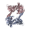
| ||||||||
| 2 | 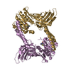
| ||||||||
| Unit cell |
|
-Components
| #1: Protein | Probability Mass: 42429.672 Da / Num. of mol.: 4 Source method: isolated from a genetically manipulated source Source: (gene. exp.) Arabidopsis thaliana (thale cress) / Gene: PRMT4.2, At1g04870, F13M7.14, F13M7_12 / Production host:  Escherichia coli (E. coli) Escherichia coli (E. coli)References: UniProt: Q9MAT5, Transferases; Transferring one-carbon groups; Methyltransferases#2: Chemical | ChemComp-SAH / S-Adenosyl-L-homocysteine  Type: L-peptide linking / Mass: 384.411 Da / Num. of mol.: 4 / Source method: obtained synthetically / Formula: C14H20N6O5S Type: L-peptide linking / Mass: 384.411 Da / Num. of mol.: 4 / Source method: obtained synthetically / Formula: C14H20N6O5S#3: Water | ChemComp-HOH / | Water Mass: 18.015 Da / Num. of mol.: 168 / Source method: isolated from a natural source / Formula: H2O Mass: 18.015 Da / Num. of mol.: 168 / Source method: isolated from a natural source / Formula: H2O |
|---|
-Experimental details
-Experiment
| Experiment | Method: X-RAY DIFFRACTION / Number of used crystals: 4 |
|---|
- Sample preparation
| Crystal | Density Matthews: 2.36 Å3/Da / Density % sol: 47.89 % |
|---|---|
| Crystal grow | Temperature: 295 K / Method: vapor diffusion, hanging drop / pH: 7.6 Details: 0.1M Tris-HCl, 2.3M Na2HPO4, 0.1M arginine, pH 7.6, VAPOR DIFFUSION, HANGING DROP, temperature 295K |
-Data collection
| Diffraction | Mean temperature: 298 K |
|---|---|
| Diffraction source | Source: ROTATING ANODE / Type: RIGAKU MICROMAX-007 HF / Wavelength: 1.5418 Å |
| Detector | Type: RIGAKU RAXIS IV++ / Detector: IMAGE PLATE / Date: Oct 10, 2009 |
| Radiation | Monochromator: VariMAX HR / Protocol: SINGLE WAVELENGTH / Monochromatic (M) / Laue (L): M / Scattering type: x-ray |
| Radiation wavelength | Wavelength: 1.5418 Å / Relative weight: 1 |
| Reflection | Resolution: 2.61→32.97 Å / Num. all: 48404 / Num. obs: 46744 / % possible obs: 96.57 % / Observed criterion σ(F): 2 / Observed criterion σ(I): 2 / Redundancy: 2.9 % / Rsym value: 0.12 |
| Reflection shell | Resolution: 2.61→2.67 Å / Redundancy: 2.6 % / Mean I/σ(I) obs: 2.3 / Rsym value: 0.58 / % possible all: 91 |
- Processing
| Software |
| |||||||||||||||||||||||||||||||||||||||||||||||||||||||||||||||||||||||||||||||||||||||||||||||||||||||||
|---|---|---|---|---|---|---|---|---|---|---|---|---|---|---|---|---|---|---|---|---|---|---|---|---|---|---|---|---|---|---|---|---|---|---|---|---|---|---|---|---|---|---|---|---|---|---|---|---|---|---|---|---|---|---|---|---|---|---|---|---|---|---|---|---|---|---|---|---|---|---|---|---|---|---|---|---|---|---|---|---|---|---|---|---|---|---|---|---|---|---|---|---|---|---|---|---|---|---|---|---|---|---|---|---|---|---|
| Refinement | Method to determine structure: MOLECULAR REPLACEMENT / Resolution: 2.61→32.97 Å / σ(F): 0 / Stereochemistry target values: TWIN_LSQ_F
| |||||||||||||||||||||||||||||||||||||||||||||||||||||||||||||||||||||||||||||||||||||||||||||||||||||||||
| Solvent computation | Shrinkage radii: 0.72 Å / VDW probe radii: 1 Å / Solvent model: FLAT BULK SOLVENT MODEL / Bsol: 30.883 Å2 / ksol: 0.312 e/Å3 | |||||||||||||||||||||||||||||||||||||||||||||||||||||||||||||||||||||||||||||||||||||||||||||||||||||||||
| Displacement parameters |
| |||||||||||||||||||||||||||||||||||||||||||||||||||||||||||||||||||||||||||||||||||||||||||||||||||||||||
| Refinement step | Cycle: LAST / Resolution: 2.61→32.97 Å
| |||||||||||||||||||||||||||||||||||||||||||||||||||||||||||||||||||||||||||||||||||||||||||||||||||||||||
| Refine LS restraints |
| |||||||||||||||||||||||||||||||||||||||||||||||||||||||||||||||||||||||||||||||||||||||||||||||||||||||||
| LS refinement shell |
|
