 Movie
Movie Controller


+ Open data

- Basic information
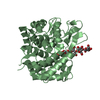
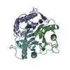
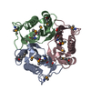
| Entry | Database: PDB / ID: 3quw | ||||||
|---|---|---|---|---|---|---|---|
| Title | Crystal structure of yeast Mmf1 | ||||||
 Components Components | Protein MMF1 | ||||||
 Keywords Keywords |  PROTEIN BINDING / chorismate mutase fold / intact mitochondria maintenance / Mitochondrial protein PROTEIN BINDING / chorismate mutase fold / intact mitochondria maintenance / Mitochondrial protein | ||||||
| Function / homology |  Function and homology information Function and homology informationorganonitrogen compound catabolic process / deaminase activity / isoleucine biosynthetic process / mitochondrial translation / mitochondrial matrix / mitochondrion / cytosolSimilarity search - Function | ||||||
| Biological species |  Saccharomyces cerevisiae (brewer's yeast) Saccharomyces cerevisiae (brewer's yeast) | ||||||
| Method | X-RAY DIFFRACTION / SYNCHROTRON / MOLECULAR REPLACEMENT / Resolution: 1.75 Å | ||||||
 Authors Authors | Jiang, Y.L. / Pu, Y.G. / Ma, X.X. / Chen, Y. / Zhou, C.Z. | ||||||
 Citation Citation | Journal: J.Struct.Biol. / Year: 2011 Title: Crystal structures and putative interface of Saccharomyces cerevisiae mitochondrial matrix proteins Mmf1 and Mam33. Authors: Pu, Y.G. / Jiang, Y.L. / Ye, X.D. / Ma, X.X. / Guo, P.C. / Lian, F.M. / Teng, Y.B. / Chen, Y. / Zhou, C.Z. | ||||||
| History |
|
- Structure visualization
| Structure viewer | Molecule: MolmilJmol/JSmol |
|---|
- Downloads & links
-Download
| PDBx/mmCIF format | 3quw.cif.gz | 38.8 KB | Display | PDBx/mmCIF format |
|---|---|---|---|---|
| PDB format | pdb3quw.ent.gz | 25.8 KB | Display | PDB format |
| PDBx/mmJSON format | 3quw.json.gz | Tree view | PDBx/mmJSON format | |
| Others |  Other downloads Other downloads |
-Validation report
| Arichive directory | https://data.pdbj.org/pub/pdb/validation_reports/qu/3quwftp://data.pdbj.org/pub/pdb/validation_reports/qu/3quw | HTTPS FTP |
|---|
-Related structure data
| Related structure data |  3qv0C 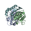 1jd1S C: citing same article ( S: Starting model for refinement |
|---|---|
| Similar structure data |


-Links
 PDBj
PDBj
- Assembly
| Deposited unit | 
| ||||||||||||
|---|---|---|---|---|---|---|---|---|---|---|---|---|---|
| 1 | 
| ||||||||||||
| Unit cell |
| ||||||||||||
| Components on special symmetry positions |
|
-Components
| #1: Protein | Mass: 16943.379 Da / Num. of mol.: 1 Source method: isolated from a genetically manipulated source Source: (gene. exp.) Saccharomyces cerevisiae (brewer's yeast)Gene: IBM1, MMF1, YIL051C / Plasmid: p28 / Production host:  Escherichia coli (E. coli) / Strain (production host): BL21 / References: UniProt: P40185 Escherichia coli (E. coli) / Strain (production host): BL21 / References: UniProt: P40185 |
|---|---|
| #2: Water | ChemComp-HOH / Water Mass: 18.015 Da / Num. of mol.: 77 / Source method: isolated from a natural source / Formula: H2O Mass: 18.015 Da / Num. of mol.: 77 / Source method: isolated from a natural source / Formula: H2O |
-Experimental details
-Experiment
| Experiment | Method: X-RAY DIFFRACTION / Number of used crystals: 1 |
|---|
- Sample preparation
| Crystal grow | Temperature: 289 K / Method: vapor diffusion, hanging drop / pH: 6.5 Details: 0.1 M Bis-Tris, 25% polyethylene glycol 3550, pH 6.5, VAPOR DIFFUSION, HANGING DROP, temperature 289K |
|---|
-Data collection
| Diffraction | Mean temperature: 100 K |
|---|---|
| Diffraction source | Source: SYNCHROTRON / Site: SSRF  / Beamline: BL17U / Wavelength: 0.97012 Å / Beamline: BL17U / Wavelength: 0.97012 Å |
| Detector | Type: RAYONIX MX-225 / Detector: CCD / Date: Dec 30, 2009 |
| Radiation | Protocol: SINGLE WAVELENGTH / Monochromatic (M) / Laue (L): M / Scattering type: x-ray |
| Radiation wavelength | Wavelength: 0.97012 Å / Relative weight: 1 |
| Reflection | Resolution: 1.74→50 Å / Num. obs: 9969 / % possible obs: 99.3 % / Observed criterion σ(F): -3 / Observed criterion σ(I): 0 / Redundancy: 11 % / Biso Wilson estimate: 29.19 Å2 / Rmerge(I) obs: 0.104 / Rsym value: 0.104 / Net I/σ(I): 14.8 |
| Reflection shell | Resolution: 1.74→1.8 Å / Redundancy: 7.8 % / Rmerge(I) obs: 0.25 / Mean I/σ(I) obs: 7.2 / Num. unique all: 929 / Rsym value: 0.25 / % possible all: 94.5 |
- Processing
| Software |
| ||||||||||||||||||||||||||||||||||||||||||||||||||||||||||||||||||||||||||||||||||||||||||||||||||||||||||||||||||||||||||||||||||||||||||||||||||||||||||||||||||||||||||
|---|---|---|---|---|---|---|---|---|---|---|---|---|---|---|---|---|---|---|---|---|---|---|---|---|---|---|---|---|---|---|---|---|---|---|---|---|---|---|---|---|---|---|---|---|---|---|---|---|---|---|---|---|---|---|---|---|---|---|---|---|---|---|---|---|---|---|---|---|---|---|---|---|---|---|---|---|---|---|---|---|---|---|---|---|---|---|---|---|---|---|---|---|---|---|---|---|---|---|---|---|---|---|---|---|---|---|---|---|---|---|---|---|---|---|---|---|---|---|---|---|---|---|---|---|---|---|---|---|---|---|---|---|---|---|---|---|---|---|---|---|---|---|---|---|---|---|---|---|---|---|---|---|---|---|---|---|---|---|---|---|---|---|---|---|---|---|---|---|---|---|---|
| Refinement | Method to determine structure: MOLECULAR REPLACEMENT Starting model: PDB entry 1JD1 Resolution: 1.75→38.81 Å / Cor.coef. Fo:Fc: 0.961 / Cor.coef. Fo:Fc free: 0.936 / SU B: 2.768 / SU ML: 0.09 / Cross valid method: THROUGHOUT / ESU R Free: 0.13 / Stereochemistry target values: MAXIMUM LIKELIHOOD / Details: HYDROGENS HAVE BEEN ADDED IN THE RIDING POSITIONS
| ||||||||||||||||||||||||||||||||||||||||||||||||||||||||||||||||||||||||||||||||||||||||||||||||||||||||||||||||||||||||||||||||||||||||||||||||||||||||||||||||||||||||||
| Solvent computation | Ion probe radii: 0.8 Å / Shrinkage radii: 0.8 Å / VDW probe radii: 1.4 Å / Solvent model: MASK | ||||||||||||||||||||||||||||||||||||||||||||||||||||||||||||||||||||||||||||||||||||||||||||||||||||||||||||||||||||||||||||||||||||||||||||||||||||||||||||||||||||||||||
| Displacement parameters | Biso mean: 22.841 Å2
| ||||||||||||||||||||||||||||||||||||||||||||||||||||||||||||||||||||||||||||||||||||||||||||||||||||||||||||||||||||||||||||||||||||||||||||||||||||||||||||||||||||||||||
| Refinement step | Cycle: LAST / Resolution: 1.75→38.81 Å
| ||||||||||||||||||||||||||||||||||||||||||||||||||||||||||||||||||||||||||||||||||||||||||||||||||||||||||||||||||||||||||||||||||||||||||||||||||||||||||||||||||||||||||
| Refine LS restraints |
| ||||||||||||||||||||||||||||||||||||||||||||||||||||||||||||||||||||||||||||||||||||||||||||||||||||||||||||||||||||||||||||||||||||||||||||||||||||||||||||||||||||||||||
| LS refinement shell | Resolution: 1.75→1.79 Å / Total num. of bins used: 20
|
