 Movie
Movie Controller


+ Open data

- Basic information
| Entry | Database: PDB / ID: 3qu3 | ||||||
|---|---|---|---|---|---|---|---|
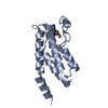
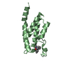
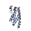
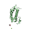
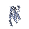
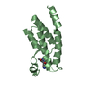
| Title | Crystal structure of IRF-7 DBD apo form | ||||||
 Components Components | Interferon regulatory factor 7 Interferon regulatory factors Interferon regulatory factors | ||||||
 Keywords Keywords | DNA BINDING PROTEIN / Helix-turn-helix / Gene regulation | ||||||
| Function / homology |  Function and homology information Function and homology informationTRAF6 mediated IRF7 activation in TLR7/8 or 9 signaling / interferon-alpha production / interferon-beta production / regulation of MyD88-independent toll-like receptor signaling pathway / TICAM1-dependent activation of IRF3/IRF7 / TRAF3-dependent IRF activation pathway / regulation of MyD88-dependent toll-like receptor signaling pathway / type I interferon production / TRAF6 mediated IRF7 activation / Activation of IRF3, IRF7 mediated by TBK1, IKKε (IKBKE) ...TRAF6 mediated IRF7 activation in TLR7/8 or 9 signaling / interferon-alpha production / interferon-beta production / regulation of MyD88-independent toll-like receptor signaling pathway / TICAM1-dependent activation of IRF3/IRF7 / TRAF3-dependent IRF activation pathway / regulation of MyD88-dependent toll-like receptor signaling pathway / type I interferon production / TRAF6 mediated IRF7 activation / Activation of IRF3, IRF7 mediated by TBK1, IKKε (IKBKE) / DEx/H-box helicases activate type I IFN and inflammatory cytokines production / regulation of adaptive immune response / positive regulation of type I interferon-mediated signaling pathway / immune system process / type I interferon-mediated signaling pathway / regulation of innate immune response / positive regulation of interferon-alpha production / immunoglobulin mediated immune response / positive regulation of type I interferon production / cis-regulatory region sequence-specific DNA binding / positive regulation of interferon-beta production / sequence-specific double-stranded DNA binding / regulation of gene expression / defense response to virus / DNA-binding transcription factor activity, RNA polymerase II-specific / RNA polymerase II cis-regulatory region sequence-specific DNA binding / positive regulation of DNA-templated transcription / positive regulation of transcription by RNA polymerase II / DNA binding / nucleoplasm / nucleus / cytosol / cytoplasmSimilarity search - Function | ||||||
| Biological species |  Mus musculus (house mouse) Mus musculus (house mouse) | ||||||
| Method | X-RAY DIFFRACTION / SYNCHROTRON / MOLECULAR REPLACEMENT / Resolution: 1.3 Å | ||||||
 Authors Authors | De Ioannes, P.E. / Escalante, C.R. / Aggarwal, A.K. | ||||||
 Citation Citation | Journal: Nucleic Acids Res. / Year: 2011 Title: Structures of apo IRF-3 and IRF-7 DNA binding domains: effect of loop L1 on DNA binding. Authors: De Ioannes, P. / Escalante, C.R. / Aggarwal, A.K. | ||||||
| History |
|
- Structure visualization
| Structure viewer | Molecule: MolmilJmol/JSmol |
|---|
- Downloads & links
-Download
| PDBx/mmCIF format | 3qu3.cif.gz | 92.8 KB | Display | PDBx/mmCIF format |
|---|---|---|---|---|
| PDB format | pdb3qu3.ent.gz | 69.5 KB | Display | PDB format |
| PDBx/mmJSON format | 3qu3.json.gz | Tree view | PDBx/mmJSON format | |
| Others |  Other downloads Other downloads |
-Validation report
| Arichive directory | https://data.pdbj.org/pub/pdb/validation_reports/qu/3qu3ftp://data.pdbj.org/pub/pdb/validation_reports/qu/3qu3 | HTTPS FTP |
|---|
-Related structure data
| Related structure data | 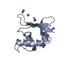 3qu6C  2o61S C: citing same article ( S: Starting model for refinement |
|---|---|
| Similar structure data |


-Links
 PDBj
PDBj

- Assembly
| Deposited unit | 
| ||||||||
|---|---|---|---|---|---|---|---|---|---|
| 1 | 
| ||||||||
| 2 | 
| ||||||||
| 3 | 
| ||||||||
| Unit cell |
|
-Components
| #1: Protein | Interferon regulatory factors / IRF-7 Mass: 15661.511 Da / Num. of mol.: 3 / Fragment: DNA Binding Domain Source method: isolated from a genetically manipulated source Source: (gene. exp.) Mus musculus (house mouse) / Gene: Irf7 / Plasmid: Pet15b / Production host:  Escherichia coli (E. coli) / Strain (production host): BL21(DE3)PLyS / References: UniProt: P70434 Escherichia coli (E. coli) / Strain (production host): BL21(DE3)PLyS / References: UniProt: P70434#2: Chemical |   Mass: 22.990 Da / Num. of mol.: 3 / Source method: obtained synthetically / Formula: Na Mass: 22.990 Da / Num. of mol.: 3 / Source method: obtained synthetically / Formula: Na#3: Chemical | ChemComp-EDO / Ethylene glycol  Mass: 62.068 Da / Num. of mol.: 4 / Source method: obtained synthetically / Formula: C2H6O2 Mass: 62.068 Da / Num. of mol.: 4 / Source method: obtained synthetically / Formula: C2H6O2#4: Water | ChemComp-HOH / | Water Mass: 18.015 Da / Num. of mol.: 287 / Source method: isolated from a natural source / Formula: H2O Mass: 18.015 Da / Num. of mol.: 287 / Source method: isolated from a natural source / Formula: H2O |
|---|
-Experimental details
-Experiment
| Experiment | Method: X-RAY DIFFRACTION / Number of used crystals: 1 |
|---|
- Sample preparation
| Crystal | Density Matthews: 1.98 Å3/Da / Density % sol: 37.97 % |
|---|---|
| Crystal grow | Temperature: 293 K Details: 19% PEG 1500, VAPOR DIFFUSION, HANGING DROP, temperature 293K |
-Data collection
| Diffraction | Mean temperature: 298 K |
|---|---|
| Diffraction source | Source: SYNCHROTRON / Site: NSLS  / Beamline: X29A / Wavelength: 1.001 / Beamline: X29A / Wavelength: 1.001 |
| Detector | Type: ADSC QUANTUM 315 / Detector: CCD / Date: Aug 8, 2008 / Details: VERTICAL FOCUSING MIRROR |
| Radiation | Monochromator: ROSENBAUM-ROCK DOUBLE CRYSTAL SAGITTAL FOCUSING MONOCHROMATOR Protocol: SINGLE WAVELENGTH / Monochromatic (M) / Laue (L): M / Scattering type: x-ray |
| Radiation wavelength | Wavelength: 1.001 Å / Relative weight: 1 |
| Reflection twin | Operator: -k,-h,-l / Fraction: 0.381 |
| Reflection | Resolution: 1.3→50 Å / Num. obs: 85736 / % possible obs: 99.8 % / Observed criterion σ(I): 1 / Redundancy: 6.5 % / Rmerge(I) obs: 0.077 / Net I/σ(I): 21.3 |
| Reflection shell | Resolution: 1.3→1.35 Å / Redundancy: 3.5 % / Rmerge(I) obs: 0.398 / Mean I/σ(I) obs: 3.4 / % possible all: 94.2 |
- Processing
| Software |
| |||||||||||||||||||||||||||||||||||||||||||||||||||||||||||||||||||||||||||||||||||||||||||||||||||||||||
|---|---|---|---|---|---|---|---|---|---|---|---|---|---|---|---|---|---|---|---|---|---|---|---|---|---|---|---|---|---|---|---|---|---|---|---|---|---|---|---|---|---|---|---|---|---|---|---|---|---|---|---|---|---|---|---|---|---|---|---|---|---|---|---|---|---|---|---|---|---|---|---|---|---|---|---|---|---|---|---|---|---|---|---|---|---|---|---|---|---|---|---|---|---|---|---|---|---|---|---|---|---|---|---|---|---|---|
| Refinement | Method to determine structure: MOLECULAR REPLACEMENT Starting model: PDB ENTRY 2O61 Resolution: 1.3→29.7 Å / σ(F): 2 / Stereochemistry target values: TWIN_LSQ_F
| |||||||||||||||||||||||||||||||||||||||||||||||||||||||||||||||||||||||||||||||||||||||||||||||||||||||||
| Solvent computation | Shrinkage radii: 0.83 Å / VDW probe radii: 1.1 Å / Solvent model: FLAT BULK SOLVENT MODEL / Bsol: 58.1 Å2 / ksol: 0.39 e/Å3 | |||||||||||||||||||||||||||||||||||||||||||||||||||||||||||||||||||||||||||||||||||||||||||||||||||||||||
| Displacement parameters | Biso mean: 22.2 Å2
| |||||||||||||||||||||||||||||||||||||||||||||||||||||||||||||||||||||||||||||||||||||||||||||||||||||||||
| Refinement step | Cycle: LAST / Resolution: 1.3→29.7 Å
| |||||||||||||||||||||||||||||||||||||||||||||||||||||||||||||||||||||||||||||||||||||||||||||||||||||||||
| Refine LS restraints |
| |||||||||||||||||||||||||||||||||||||||||||||||||||||||||||||||||||||||||||||||||||||||||||||||||||||||||
| LS refinement shell |
|
