 Movie
Movie Controller

[English] 日本語
 Yorodumi
- PDB-3qtt: Crystal Structure of Pantoate-beta-alanine Ligase from Francisell... -
+ Open data

- Basic information
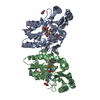
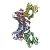
| Entry | Database: PDB / ID: 3qtt | ||||||
|---|---|---|---|---|---|---|---|
| Title | Crystal Structure of Pantoate-beta-alanine Ligase from Francisella tularensis Complexed with Beta-gamma ATP and Beta-alanine | ||||||
 Components Components | Pantothenate synthetase | ||||||
 Keywords Keywords |  LIGASE / Structural Genomics / Center for Structural Genomics of Infectious Diseases / CSGID / alpha-beta sandwich / cytosol LIGASE / Structural Genomics / Center for Structural Genomics of Infectious Diseases / CSGID / alpha-beta sandwich / cytosol | ||||||
| Function / homology |  Function and homology information Function and homology informationpantoate-beta-alanine ligase (AMP-forming) / pantoate-beta-alanine ligase activity / pantothenate biosynthetic process / ATP binding / cytosolSimilarity search - Function | ||||||
| Biological species |  Francisella tularensis subsp. tularensis (bacteria) Francisella tularensis subsp. tularensis (bacteria) | ||||||
| Method | X-RAY DIFFRACTION / SYNCHROTRON / SAD / Resolution: 2.599 Å | ||||||
 Authors Authors | Maltseva, N. / Kim, Y. / Kwon, K. / Anderson, W.F. / Joachimiak, A. / Center for Structural Genomics of Infectious Diseases (CSGID) | ||||||
 Citation Citation | Journal: To be Published Title: Crystal Structure of Pantoate-beta-alanine Ligase from Francisella tularensis Complexed with Beta-gamma ATP and Beta-alanine. Authors: Maltseva, N. / Kim, Y. / Kwon, K. / Anderson, W.F. / Joachimiak, A. | ||||||
| History |
|
- Structure visualization
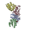
| Structure viewer | Molecule: MolmilJmol/JSmol |
|---|
- Downloads & links
-Download
| PDBx/mmCIF format | 3qtt.cif.gz | 228.4 KB | Display | PDBx/mmCIF format |
|---|---|---|---|---|
| PDB format | pdb3qtt.ent.gz | 193.9 KB | Display | PDB format |
| PDBx/mmJSON format | 3qtt.json.gz | Tree view | PDBx/mmJSON format | |
| Others |  Other downloads Other downloads |
-Validation report
| Arichive directory | https://data.pdbj.org/pub/pdb/validation_reports/qt/3qttftp://data.pdbj.org/pub/pdb/validation_reports/qt/3qtt | HTTPS FTP |
|---|
-Related structure data
| Related structure data | |
|---|---|
| Similar structure data | |
| Other databases |


-Links
 PDBj
PDBj


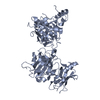
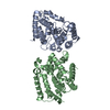
- Assembly
| Deposited unit | 
| ||||||||
|---|---|---|---|---|---|---|---|---|---|
| 1 | 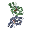
| ||||||||
| 2 | 
| ||||||||
| 3 | 
| ||||||||
| Unit cell |
|
-Components
-Protein , 1 types, 2 molecules AB
| #1: Protein | Mass: 30209.178 Da / Num. of mol.: 2 Source method: isolated from a genetically manipulated source Source: (gene. exp.) Francisella tularensis subsp. tularensis (bacteria)Strain: SCHU S4 / Gene: FTT_1390, panC / Plasmid: pMCSG7 / Production host: Escherichia coli (E. coli) / Strain (production host): BL21 magicReferences: UniProt: Q5NF57, pantoate-beta-alanine ligase (AMP-forming) |
|---|
-Non-polymers , 5 types, 113 molecules 


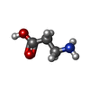

| #2: Chemical |  Mass: 506.196 Da / Num. of mol.: 2 / Source method: obtained synthetically / Formula: C10H17N6O12P3 / Comment: AMP-PNP, energy-carrying molecule analogue*YM Mass: 506.196 Da / Num. of mol.: 2 / Source method: obtained synthetically / Formula: C10H17N6O12P3 / Comment: AMP-PNP, energy-carrying molecule analogue*YM#3: Chemical | Proline Type: L-peptide linking / Mass: 115.130 Da / Num. of mol.: 2 / Source method: obtained synthetically / Formula: C5H9NO2 Type: L-peptide linking / Mass: 115.130 Da / Num. of mol.: 2 / Source method: obtained synthetically / Formula: C5H9NO2#4: Chemical | ChemComp-MG / |  Mass: 24.305 Da / Num. of mol.: 1 / Source method: obtained synthetically / Formula: Mg Mass: 24.305 Da / Num. of mol.: 1 / Source method: obtained synthetically / Formula: Mg#5: Chemical | Β-Alanine Type: peptide-like / Mass: 89.093 Da / Num. of mol.: 2 / Source method: obtained synthetically / Formula: C3H7NO2 Type: peptide-like / Mass: 89.093 Da / Num. of mol.: 2 / Source method: obtained synthetically / Formula: C3H7NO2#6: Water | ChemComp-HOH / | WaterMass: 18.015 Da / Num. of mol.: 106 / Source method: isolated from a natural source / Formula: H2O |
|---|
-Experimental details
-Experiment
| Experiment | Method: X-RAY DIFFRACTION / Number of used crystals: 1 |
|---|
- Sample preparation
| Crystal | Density Matthews: 2.37 Å3/Da / Density % sol: 48.1 % |
|---|---|
| Crystal grow | Temperature: 289 K / Method: vapor diffusion, sitting drop / pH: 7.5 Details: 0.2 M Proline, 0.1 M HEPES pH 7.5, 10 % w/v PEG 3350, VAPOR DIFFUSION, SITTING DROP, temperature 289K |
-Data collection
| Diffraction | Mean temperature: 100 K |
|---|---|
| Diffraction source | Source: SYNCHROTRON / Site: APS  / Beamline: 19-ID / Wavelength: 0.97912 Å / Beamline: 19-ID / Wavelength: 0.97912 Å |
| Detector | Type: ADSC QUANTUM 315r / Detector: CCD / Date: Dec 9, 2010 / Details: mirrors |
| Radiation | Monochromator: double crystal monochromator / Protocol: SINGLE WAVELENGTH / Monochromatic (M) / Laue (L): M / Scattering type: x-ray |
| Radiation wavelength | Wavelength: 0.97912 Å / Relative weight: 1 |
| Reflection | Resolution: 2.6→50 Å / Num. all: 17255 / Num. obs: 17255 / % possible obs: 99.8 % / Observed criterion σ(F): 0 / Observed criterion σ(I): 0 / Redundancy: 7.6 % / Rsym value: 0.091 / Net I/σ(I): 9 |
| Reflection shell | Resolution: 2.6→2.64 Å / Redundancy: 7.6 % / Mean I/σ(I) obs: 3.1 / Rsym value: 0.759 / % possible all: 99.8 |
- Processing
| Software |
| |||||||||||||||||||||||||||||||||||||||||||||||||||||||||||||||||||||||||||||||||||||||||||
|---|---|---|---|---|---|---|---|---|---|---|---|---|---|---|---|---|---|---|---|---|---|---|---|---|---|---|---|---|---|---|---|---|---|---|---|---|---|---|---|---|---|---|---|---|---|---|---|---|---|---|---|---|---|---|---|---|---|---|---|---|---|---|---|---|---|---|---|---|---|---|---|---|---|---|---|---|---|---|---|---|---|---|---|---|---|---|---|---|---|---|---|---|
| Refinement | Method to determine structure: SAD / Resolution: 2.599→33.338 Å / SU ML: 0.43 / Isotropic thermal model: mixed / σ(F): 0.4 / Phase error: 28.37 / Stereochemistry target values: MLHL
| |||||||||||||||||||||||||||||||||||||||||||||||||||||||||||||||||||||||||||||||||||||||||||
| Solvent computation | Shrinkage radii: 0.95 Å / VDW probe radii: 1.2 Å / Solvent model: FLAT BULK SOLVENT MODEL / Bsol: 64.732 Å2 / ksol: 0.304 e/Å3 | |||||||||||||||||||||||||||||||||||||||||||||||||||||||||||||||||||||||||||||||||||||||||||
| Displacement parameters |
| |||||||||||||||||||||||||||||||||||||||||||||||||||||||||||||||||||||||||||||||||||||||||||
| Refinement step | Cycle: LAST / Resolution: 2.599→33.338 Å
| |||||||||||||||||||||||||||||||||||||||||||||||||||||||||||||||||||||||||||||||||||||||||||
| Refine LS restraints |
| |||||||||||||||||||||||||||||||||||||||||||||||||||||||||||||||||||||||||||||||||||||||||||
| LS refinement shell |
| |||||||||||||||||||||||||||||||||||||||||||||||||||||||||||||||||||||||||||||||||||||||||||
| Refinement TLS params. | Method: refined / Refine-ID: X-RAY DIFFRACTION
| |||||||||||||||||||||||||||||||||||||||||||||||||||||||||||||||||||||||||||||||||||||||||||
| Refinement TLS group |
|
