Monochromator: double crystal / Protocol: SINGLE WAVELENGTH / Monochromatic (M) / Laue (L): M / Scattering type: x-ray
Radiation wavelength
Wavelength: 0.97926 Å / Relative weight: 1
Reflection
Resolution: 1.9→50 Å / Num. all: 25141 / Num. obs: 25034 / % possible obs: 99.6 % / Observed criterion σ(I): -3 / Redundancy: 3.7 % / Biso Wilson estimate: 35.41 Å2 / Rmerge(I) obs: 0.065 / Net I/σ(I): 25.1
Reflection shell
Resolution: 1.9→1.93 Å / Redundancy: 3.5 % / Rmerge(I) obs: 0.421 / Mean I/σ(I) obs: 2.5 / % possible all: 99.4
-
Processing
Software
Name
Version
Classification
SBC-Collect
datacollection
PHASER
phasing
PHENIX
refinement
BUSTER
2.8.0
refinement
HKL-3000
datareduction
HKL-3000
datascaling
Refinement
Method to determine structure: MOLECULAR REPLACEMENT Starting model: different crystal form of the same protein (solved by SAD) Resolution: 1.9→21.99 Å / Cor.coef. Fo:Fc: 0.9518 / Cor.coef. Fo:Fc free: 0.9454 / Isotropic thermal model: isotropic / Cross valid method: THROUGHOUT / σ(F): 0
Rfactor
Num. reflection
% reflection
Selection details
Rfree
0.201
1271
5.08 %
RANDOM
Rwork
0.185
-
-
-
obs
0.1859
25015
-
-
all
-
25015
-
-
Displacement parameters
Biso mean: 49.09 Å2
Baniso -1
Baniso -2
Baniso -3
1-
3.6729 Å2
0 Å2
-1.5544 Å2
2-
-
4.485 Å2
0 Å2
3-
-
-
-8.1579 Å2
Refine analyze
Luzzati coordinate error obs: 0.275 Å
Refinement step
Cycle: LAST / Resolution: 1.9→21.99 Å
Protein
Nucleic acid
Ligand
Solvent
Total
Num. atoms
2095
0
2
74
2171
Refine LS restraints
Refine-ID
Type
Dev ideal
Number
Restraint function
Weight
X-RAY DIFFRACTION
t_bond_d
0.012
2146
HARMONIC
2
X-RAY DIFFRACTION
t_angle_deg
1.09
2911
HARMONIC
2
X-RAY DIFFRACTION
t_dihedral_angle_d
734
SINUSOIDAL
2
X-RAY DIFFRACTION
t_incorr_chiral_ct
X-RAY DIFFRACTION
t_pseud_angle
X-RAY DIFFRACTION
t_trig_c_planes
44
HARMONIC
2
X-RAY DIFFRACTION
t_gen_planes
332
HARMONIC
5
X-RAY DIFFRACTION
t_it
2146
HARMONIC
20
X-RAY DIFFRACTION
t_nbd
0
SEMIHARMONIC
5
X-RAY DIFFRACTION
t_omega_torsion
3.3
X-RAY DIFFRACTION
t_other_torsion
16.47
X-RAY DIFFRACTION
t_improper_torsion
X-RAY DIFFRACTION
t_chiral_improper_torsion
276
SEMIHARMONIC
5
X-RAY DIFFRACTION
t_sum_occupancies
X-RAY DIFFRACTION
t_utility_distance
X-RAY DIFFRACTION
t_utility_angle
X-RAY DIFFRACTION
t_utility_torsion
X-RAY DIFFRACTION
t_ideal_dist_contact
2521
SEMIHARMONIC
4
LS refinement shell
Resolution: 1.9→1.98 Å / Total num. of bins used: 13
Rfactor
Num. reflection
% reflection
Rfree
0.2286
128
4.61 %
Rwork
0.1985
2650
-
all
0.1998
2778
-
Refinement TLS params.
Method: refined / Refine-ID: X-RAY DIFFRACTION
ID
L11 (°2)
L12 (°2)
L13 (°2)
L22 (°2)
L23 (°2)
L33 (°2)
S11 (Å °)
S12 (Å °)
S13 (Å °)
S21 (Å °)
S22 (Å °)
S23 (Å °)
S31 (Å °)
S32 (Å °)
S33 (Å °)
T11 (Å2)
T12 (Å2)
T13 (Å2)
T22 (Å2)
T23 (Å2)
T33 (Å2)
Origin x (Å)
Origin y (Å)
Origin z (Å)
1
2.2555
-2.1859
2.6265
0.7054
-1.3893
5.2078
-0.0057
-0.2965
0.0362
0.7017
-0.0729
-0.2801
0.0757
0.238
0.0786
0.1064
-0.0008
-0.1724
-0.191
0.0082
0.0245
31.2748
16.2349
15.9574
2
4.7423
2.1978
3.3214
3.4903
-0.0752
6.2713
0.0027
-0.6781
0.0917
0.567
-0.0274
-0.3467
-0.0675
0.2797
0.0247
-0.0705
0.0057
-0.3045
-0.304
-0.0195
0.118
35.2535
23.8196
19.709
3
2.173
-0.6067
0.1478
5.446
-0.2317
1.3652
-0.0584
-0.0076
0.0221
0.2276
-0.0413
-0.4949
-0.086
0.0428
0.0997
-0.1597
-0.0041
-0.0232
-0.1501
0.0018
0.1111
30.4746
21.18
2.7378
4
6.206
-0.947
-1.7518
0
0.594
1.4625
-0.1276
-0.6445
-0.7011
0.1176
0.0289
0.0133
0.1538
0.1397
0.0987
-0.062
0.0154
-0.007
-0.0906
0.0807
0.0786
-0.7016
16.6032
6.1986
5
5.711
0.0867
-2.3143
0.463
0.4974
2.1195
0.2346
-0.6871
0.4274
0.1466
-0.0018
-0.1448
-0.1271
0.26
-0.2328
-0.1685
-0.0254
-0.0116
-0.0797
-0.0477
0.006
6.3413
30.0827
5.6762
6
-0.2369
-3.1057
-0.3528
0.595
-0.0905
3.1604
0.0178
-0.3369
-0.0619
0.3546
-0.1448
0.0467
0.2002
-0.1315
0.127
-0.1183
-0.0813
-0.0452
0.1351
0.0746
-0.2986
-6.2083
21.381
19.991
Refinement TLS group
ID
Refine-ID
Refine TLS-ID
Selection details
Auth asym-ID
Auth seq-ID
1
X-RAY DIFFRACTION
1
{A|1 - A|23}
A
1 - 23
2
X-RAY DIFFRACTION
2
{A|24 - A|61}
A
24 - 61
3
X-RAY DIFFRACTION
3
{A|62 - A|163}
A
62 - 163
4
X-RAY DIFFRACTION
4
{A|164 - A|224}
A
164 - 224
5
X-RAY DIFFRACTION
5
{A|231 - A|271}
A
231 - 271
6
X-RAY DIFFRACTION
6
{A|274 - A|285}
A
274 - 285
+
About Yorodumi
-
News
-
Feb 9, 2022. New format data for meta-information of EMDB entries
New format data for meta-information of EMDB entries
Version 3 of the EMDB header file is now the official format.
The previous official version 1.9 will be removed from the archive.
In the structure databanks used in Yorodumi, some data are registered as the other names, "COVID-19 virus" and "2019-nCoV". Here are the details of the virus and the list of structure data.
Jan 31, 2019. EMDB accession codes are about to change! (news from PDBe EMDB page)
EMDB accession codes are about to change! (news from PDBe EMDB page)
The allocation of 4 digits for EMDB accession codes will soon come to an end. Whilst these codes will remain in use, new EMDB accession codes will include an additional digit and will expand incrementally as the available range of codes is exhausted. The current 4-digit format prefixed with “EMD-” (i.e. EMD-XXXX) will advance to a 5-digit format (i.e. EMD-XXXXX), and so on. It is currently estimated that the 4-digit codes will be depleted around Spring 2019, at which point the 5-digit format will come into force.
The EM Navigator/Yorodumi systems omit the EMD- prefix.
Related info.:Q: What is EMD? / ID/Accession-code notation in Yorodumi/EM Navigator
Yorodumi is a browser for structure data from EMDB, PDB, SASBDB, etc.
This page is also the successor to EM Navigator detail page, and also detail information page/front-end page for Omokage search.
The word "yorodu" (or yorozu) is an old Japanese word meaning "ten thousand". "mi" (miru) is to see.
Related info.:EMDB / PDB / SASBDB / Comparison of 3 databanks / Yorodumi Search / Aug 31, 2016. New EM Navigator & Yorodumi / Yorodumi Papers / Jmol/JSmol / Function and homology information / Changes in new EM Navigator and Yorodumi
 Movie
Movie Controller

 Yorodumi
 Open data
 Basic information
 Components
 Keywords
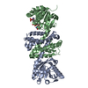
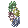
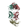
 Structural genomics / Unknown function / PSI-Biology / Midwest Center for Structural Genomics / MCSG / Rossmann-fold NAD-binding proteins
 Function and homology information
 Authors
 Citation
 Structure visualization
 Downloads & links
 Other downloads
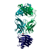


 PDBj
PDBj


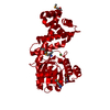
 Assembly
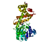

 Mass: 35.453 Da / Num. of mol.: 2 / Source method: obtained synthetically / Formula: Cl
 Mass: 18.015 Da / Num. of mol.: 74 / Source method: isolated from a natural source / Formula: H2O
 Sample preparation
 / Beamline: 19-ID / Wavelength: 0.97926 Å
 Processing