Type: MARMOSAIC 325 mm CCD / Detector: CCD / Date: Mar 21, 2009
Radiation
Monochromator: Double crystal monochromator / Protocol: SINGLE WAVELENGTH / Monochromatic (M) / Laue (L): M / Scattering type: x-ray
Radiation wavelength
Wavelength: 0.97915 Å / Relative weight: 1
Reflection
Redundancy: 7.8 % / Av σ(I) over netI: 18.35 / Number: 290928 / Rmerge(I) obs: 0.084 / Χ2: 1.06 / D res high: 2.1 Å / D res low: 40 Å / Num. obs: 37509 / % possible obs: 97.6
Diffraction reflection shell
Highest resolution (Å)
Lowest resolution (Å)
% possible obs (%)
ID
Rmerge(I) obs
Chi squared
Redundancy
4.52
40
99.7
1
0.05
1.066
11
3.59
4.52
100
1
0.069
1.065
10.4
3.14
3.59
100
1
0.091
0.988
9.8
2.85
3.14
100
1
0.136
0.996
9.4
2.65
2.85
100
1
0.198
1.086
8.8
2.49
2.65
100
1
0.259
1.006
7.8
2.37
2.49
100
1
0.321
1.083
6.6
2.26
2.37
99.3
1
0.411
1.28
5.4
2.18
2.26
95
1
0.472
1.093
4.2
2.1
2.18
81.8
1
0.514
1.242
3.1
Reflection
Resolution: 2.1→40 Å / Num. obs: 37509 / % possible obs: 97.6 % / Redundancy: 7.8 % / Rmerge(I) obs: 0.084 / Χ2: 1.064 / Net I/σ(I): 15
Reflection shell
Resolution (Å)
Redundancy (%)
Rmerge(I) obs
Num. unique all
Χ2
% possible all
2.1-2.18
3.1
0.514
3141
1.242
81.8
2.18-2.26
4.2
0.472
3674
1.093
95
2.26-2.37
5.4
0.411
3755
1.28
99.3
2.37-2.49
6.6
0.321
3858
1.083
100
2.49-2.65
7.8
0.259
3823
1.006
100
2.65-2.85
8.8
0.198
3815
1.086
100
2.85-3.14
9.4
0.136
3843
0.996
100
3.14-3.59
9.8
0.091
3847
0.988
100
3.59-4.52
10.4
0.069
3860
1.065
100
4.52-40
11
0.05
3893
1.066
99.7
-
Phasing
Phasing
Method: molecular replacement
Phasing MR
Model details: Phaser MODE: MR_AUTO
Highest resolution
Lowest resolution
Rotation
2.5 Å
33.15 Å
Translation
2.5 Å
33.15 Å
-
Processing
Software
Name
Version
Classification
NB
DENZO
datareduction
SCALEPACK
datascaling
PHASER
1.3.3
phasing
REFMAC
refinement
PDB_EXTRACT
3.1
dataextraction
HKL-2000
datacollection
HKL-2000
datareduction
HKL-2000
datascaling
Refinement
Method to determine structure: MOLECULAR REPLACEMENT / Resolution: 2.1→33 Å / Cor.coef. Fo:Fc: 0.96 / Cor.coef. Fo:Fc free: 0.941 / WRfactor Rfree: 0.2434 / WRfactor Rwork: 0.1984 / Occupancy max: 1 / Occupancy min: 0.33 / FOM work R set: 0.8242 / SU B: 9.885 / SU ML: 0.138 / SU R Cruickshank DPI: 0.2027 / SU Rfree: 0.1781 / Cross valid method: THROUGHOUT / σ(F): 0 / ESU R Free: 0.179 / Stereochemistry target values: MAXIMUM LIKELIHOOD / Details: HYDROGENS HAVE BEEN ADDED IN THE RIDING POSITIONS
Rfactor
Num. reflection
% reflection
Selection details
Rfree
0.2349
1874
5 %
RANDOM
Rwork
0.189
-
-
-
obs
0.1913
37441
97.49 %
-
Solvent computation
Ion probe radii: 0.8 Å / Shrinkage radii: 0.8 Å / VDW probe radii: 1.2 Å / Solvent model: MASK
In the structure databanks used in Yorodumi, some data are registered as the other names, "COVID-19 virus" and "2019-nCoV". Here are the details of the virus and the list of structure data.
Jan 31, 2019. EMDB accession codes are about to change! (news from PDBe EMDB page)
EMDB accession codes are about to change! (news from PDBe EMDB page)
The allocation of 4 digits for EMDB accession codes will soon come to an end. Whilst these codes will remain in use, new EMDB accession codes will include an additional digit and will expand incrementally as the available range of codes is exhausted. The current 4-digit format prefixed with “EMD-” (i.e. EMD-XXXX) will advance to a 5-digit format (i.e. EMD-XXXXX), and so on. It is currently estimated that the 4-digit codes will be depleted around Spring 2019, at which point the 5-digit format will come into force.
The EM Navigator/Yorodumi systems omit the EMD- prefix.
Related info.:Q: What is EMD? / ID/Accession-code notation in Yorodumi/EM Navigator
Yorodumi is a browser for structure data from EMDB, PDB, SASBDB, etc.
This page is also the successor to EM Navigator detail page, and also detail information page/front-end page for Omokage search.
The word "yorodu" (or yorozu) is an old Japanese word meaning "ten thousand". "mi" (miru) is to see.
Related info.:EMDB / PDB / SASBDB / Comparison of 3 databanks / Yorodumi Search / Aug 31, 2016. New EM Navigator & Yorodumi / Yorodumi Papers / Jmol/JSmol / Function and homology information / Changes in new EM Navigator and Yorodumi
 Movie
Movie Controller

 Yorodumi
 Open data
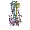
 Basic information
 Components
 ) x 2
) x 2  Keywords
 Function and homology information

 Authors
 Citation
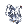
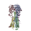
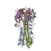
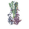
 Structure visualization
 Downloads & links
 Other downloads
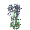


 PDBj
PDBj


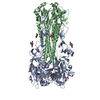
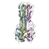
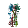
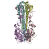
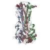
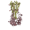
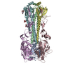
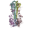
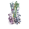
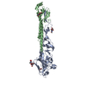
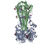
 Assembly


 Type: D-saccharide, beta linking / Mass: 221.208 Da / Num. of mol.: 2


 Mass: 62.068 Da / Num. of mol.: 1 / Source method: obtained synthetically / Formula: C2H6O2
 Mass: 106.120 Da / Num. of mol.: 1 / Source method: obtained synthetically / Formula: C4H10O3
 Sample preparation
 / Beamline: BL9-2 / Wavelength: 0.97915 Å
 Processing