 Movie
Movie Controller

[English] 日本語
 Yorodumi
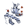
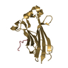
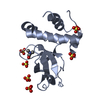
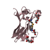
- PDB-3qkj: The PWWP domain of human DNA (CYTOSINE-5-)-METHYLTRANSFERASE 3 BE... -
+ Open data

- Basic information
| Entry | Database: PDB / ID: 3qkj | ||||||
|---|---|---|---|---|---|---|---|
| Title | The PWWP domain of human DNA (CYTOSINE-5-)-METHYLTRANSFERASE 3 BETA in complex with a bis-tris molecule | ||||||
 Components Components | DNA cytosine-5 methyltransferase 3 beta isoform 6 variant | ||||||
 Keywords Keywords |  TRANSFERASE / DNMT3B / PWWP DOMAIN / METHYLTRANSFERASE 3 BETA / SGC / S-ADENOSYL-L-METHIONINE / ZINC-FINGER / Structural Genomics / Structural Genomics Consortium TRANSFERASE / DNMT3B / PWWP DOMAIN / METHYLTRANSFERASE 3 BETA / SGC / S-ADENOSYL-L-METHIONINE / ZINC-FINGER / Structural Genomics / Structural Genomics Consortium | ||||||
| Function / homology |  Function and homology information Function and homology information: / DNA (cytosine-5-)-methyltransferase activity, acting on CpG substrates / DNA-methyltransferase activity / DNA (cytosine-5-)-methyltransferase / DNA (cytosine-5-)-methyltransferase activity / SUMOylation of DNA methylation proteins / : / catalytic complex / DNA methylation / PRC2 methylates histones and DNA ...: / DNA (cytosine-5-)-methyltransferase activity, acting on CpG substrates / DNA-methyltransferase activity / DNA (cytosine-5-)-methyltransferase / DNA (cytosine-5-)-methyltransferase activity / SUMOylation of DNA methylation proteins / : / catalytic complex / DNA methylation / PRC2 methylates histones and DNA / methyltransferase activity / Defective pyroptosis / NoRC negatively regulates rRNA expression / transcription corepressor activity / methylation / positive regulation of gene expression / negative regulation of transcription by RNA polymerase II / DNA binding / nucleoplasm / metal ion binding / nucleusSimilarity search - Function | ||||||
| Biological species |  Homo sapiens (human) Homo sapiens (human) | ||||||
| Method | X-RAY DIFFRACTION / SYNCHROTRON / MOLECULAR REPLACEMENT / Resolution: 2.04 Å | ||||||
 Authors Authors | Zeng, H. / Amaya, M.F. / Mackenzie, F. / Weigelt, J. / Sundstrom, M. / Arrowsmith, C.H. / Edwards, A.M. / Botchkarev, A. / Min, J. / Wu, H. / Structural Genomics Consortium (SGC) | ||||||
 Citation Citation | Journal: Plos One / Year: 2011 Title: Structural and Histone Binding Ability Characterizations of Human PWWP Domains. Authors: Wu, H. / Zeng, H. / Lam, R. / Tempel, W. / Amaya, M.F. / Xu, C. / Dombrovski, L. / Qiu, W. / Wang, Y. / Min, J. | ||||||
| History |
|
- Structure visualization
| Structure viewer | Molecule: MolmilJmol/JSmol |
|---|
- Downloads & links
-Download
| PDBx/mmCIF format | 3qkj.cif.gz | 224.5 KB | Display | PDBx/mmCIF format |
|---|---|---|---|---|
| PDB format | pdb3qkj.ent.gz | 183.8 KB | Display | PDB format |
| PDBx/mmJSON format | 3qkj.json.gz | Tree view | PDBx/mmJSON format | |
| Others |  Other downloads Other downloads |
-Validation report
| Arichive directory | https://data.pdbj.org/pub/pdb/validation_reports/qk/3qkjftp://data.pdbj.org/pub/pdb/validation_reports/qk/3qkj | HTTPS FTP |
|---|
-Related structure data
| Related structure data |  3eaeC 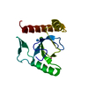 3l42C 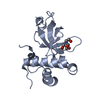 3llrC 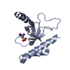 3lyiC 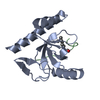 3mo8C 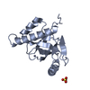 3pfsC 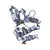 3pmiC  3qbyC  3qj6C C: citing same article ( |
|---|---|
| Similar structure data |


-Links
 PDBj
PDBj


- Assembly
| Deposited unit | 
| ||||||||
|---|---|---|---|---|---|---|---|---|---|
| 1 | 
| ||||||||
| 2 | 
| ||||||||
| 3 | 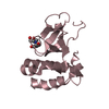
| ||||||||
| 4 | 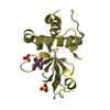
| ||||||||
| Unit cell |
|
-Components
| #1: Protein | Mass: 16755.896 Da / Num. of mol.: 4 / Fragment: UNP residues 293-442 Source method: isolated from a genetically manipulated source Source: (gene. exp.) Homo sapiens (human) / Plasmid: pET28-MHL / Production host:  Escherichia coli (E. coli) / Strain (production host): BL21(DE3) / References: UniProt: Q59H79, UniProt: Q9UBC3*PLUS Escherichia coli (E. coli) / Strain (production host): BL21(DE3) / References: UniProt: Q59H79, UniProt: Q9UBC3*PLUS#2: Chemical | ChemComp-BTB / Bis-tris methane  Mass: 209.240 Da / Num. of mol.: 4 / Source method: obtained synthetically / Formula: C8H19NO5 / Comment: pH buffer*YM Mass: 209.240 Da / Num. of mol.: 4 / Source method: obtained synthetically / Formula: C8H19NO5 / Comment: pH buffer*YM#3: Chemical | ChemComp-SO4 / Sulfate  Mass: 96.063 Da / Num. of mol.: 6 / Source method: obtained synthetically / Formula: SO4 Mass: 96.063 Da / Num. of mol.: 6 / Source method: obtained synthetically / Formula: SO4#4: Water | ChemComp-HOH / | Water Mass: 18.015 Da / Num. of mol.: 263 / Source method: isolated from a natural source / Formula: H2O Mass: 18.015 Da / Num. of mol.: 263 / Source method: isolated from a natural source / Formula: H2O |
|---|
-Experimental details
-Experiment
| Experiment | Method: X-RAY DIFFRACTION / Number of used crystals: 1 |
|---|
- Sample preparation
| Crystal | Density Matthews: 3.84 Å3/Da / Density % sol: 67.93 % |
|---|---|
| Crystal grow | Temperature: 300 K / Method: vapor diffusion, sitting drop / pH: 6 Details: pH 6, VAPOR DIFFUSION, SITTING DROP, temperature 300K |
-Data collection
| Diffraction | Mean temperature: 100 K |
|---|---|
| Diffraction source | Source: SYNCHROTRON / Site: NSLS  / Beamline: X12B / Wavelength: 1 Å / Beamline: X12B / Wavelength: 1 Å |
| Detector | Type: ADSC QUANTUM 210 / Detector: CCD / Date: Jun 5, 2009 |
| Radiation | Monochromator: si / Protocol: SINGLE WAVELENGTH / Monochromatic (M) / Laue (L): M / Scattering type: x-ray |
| Radiation wavelength | Wavelength: 1 Å / Relative weight: 1 |
| Reflection | Resolution: 2.04→50 Å / Num. obs: 62267 / % possible obs: 98.9 % / Observed criterion σ(F): 0 / Observed criterion σ(I): 0 / Biso Wilson estimate: 51.72 Å2 |
- Processing
| Software |
| |||||||||||||||||||||||||||||||||||||||||||||||||||||||||||||||||||||||||||||||||||||||||||||||||||||||||||||||||||||||||||||
|---|---|---|---|---|---|---|---|---|---|---|---|---|---|---|---|---|---|---|---|---|---|---|---|---|---|---|---|---|---|---|---|---|---|---|---|---|---|---|---|---|---|---|---|---|---|---|---|---|---|---|---|---|---|---|---|---|---|---|---|---|---|---|---|---|---|---|---|---|---|---|---|---|---|---|---|---|---|---|---|---|---|---|---|---|---|---|---|---|---|---|---|---|---|---|---|---|---|---|---|---|---|---|---|---|---|---|---|---|---|---|---|---|---|---|---|---|---|---|---|---|---|---|---|---|---|---|
| Refinement | Method to determine structure: MOLECULAR REPLACEMENT / Resolution: 2.04→34.04 Å / Cor.coef. Fo:Fc: 0.9567 / Cor.coef. Fo:Fc free: 0.9454 / SU B: 4.788 / SU ML: 0.129 / Cross valid method: THROUGHOUT / σ(F): 0 / ESU R Free: 0.158 / Stereochemistry target values: MAXIMUM LIKELIHOOD / Details: HYDROGENS HAVE BEEN ADDED IN THE RIDING POSITIONS
| |||||||||||||||||||||||||||||||||||||||||||||||||||||||||||||||||||||||||||||||||||||||||||||||||||||||||||||||||||||||||||||
| Solvent computation | Ion probe radii: 0.8 Å / Shrinkage radii: 0.8 Å / VDW probe radii: 1.4 Å / Solvent model: MASK | |||||||||||||||||||||||||||||||||||||||||||||||||||||||||||||||||||||||||||||||||||||||||||||||||||||||||||||||||||||||||||||
| Displacement parameters | Biso mean: 59.37 Å2
| |||||||||||||||||||||||||||||||||||||||||||||||||||||||||||||||||||||||||||||||||||||||||||||||||||||||||||||||||||||||||||||
| Refine analyze | Luzzati coordinate error obs: 0.39 Å | |||||||||||||||||||||||||||||||||||||||||||||||||||||||||||||||||||||||||||||||||||||||||||||||||||||||||||||||||||||||||||||
| Refinement step | Cycle: LAST / Resolution: 2.04→34.04 Å
| |||||||||||||||||||||||||||||||||||||||||||||||||||||||||||||||||||||||||||||||||||||||||||||||||||||||||||||||||||||||||||||
| Refine LS restraints |
| |||||||||||||||||||||||||||||||||||||||||||||||||||||||||||||||||||||||||||||||||||||||||||||||||||||||||||||||||||||||||||||
| LS refinement shell | Resolution: 2.04→2.09 Å / Total num. of bins used: 20
| |||||||||||||||||||||||||||||||||||||||||||||||||||||||||||||||||||||||||||||||||||||||||||||||||||||||||||||||||||||||||||||
| Refinement TLS params. | Method: refined / Refine-ID: X-RAY DIFFRACTION
| |||||||||||||||||||||||||||||||||||||||||||||||||||||||||||||||||||||||||||||||||||||||||||||||||||||||||||||||||||||||||||||
| Refinement TLS group |
|
