 Movie
Movie Controller


+ Open data

- Basic information
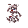
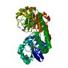
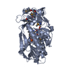
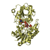
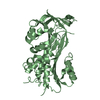
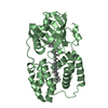
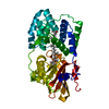
| Entry | Database: PDB / ID: 3qj4 | ||||||
|---|---|---|---|---|---|---|---|
| Title | Crystal structure of Human Renalase (isoform 1) | ||||||
 Components Components | Renalase | ||||||
 Keywords Keywords | OXIDOREDUCTASE / FAD/NAD(P)-binding Rossmann fold Superfamily / Flavin containing amine oxidoreductase / Monoamine oxidase / NAD / extracellular | ||||||
| Function / homology |  Function and homology informationrenalase / epinephrine binding / monoamine oxidase activity / response to epinephrine / NAD biosynthesis via nicotinamide riboside salvage pathway / NADH binding / : / response to salt / Nicotinamide salvaging / oxidoreductase activity, acting on NAD(P)H ...renalase / epinephrine binding / monoamine oxidase activity / response to epinephrine / NAD biosynthesis via nicotinamide riboside salvage pathway / NADH binding / : / response to salt / Nicotinamide salvaging / oxidoreductase activity, acting on NAD(P)H / negative regulation of heart rate / negative regulation of blood pressure / response to ischemia / extracellular space / extracellular region Function and homology informationrenalase / epinephrine binding / monoamine oxidase activity / response to epinephrine / NAD biosynthesis via nicotinamide riboside salvage pathway / NADH binding / : / response to salt / Nicotinamide salvaging / oxidoreductase activity, acting on NAD(P)H ...renalase / epinephrine binding / monoamine oxidase activity / response to epinephrine / NAD biosynthesis via nicotinamide riboside salvage pathway / NADH binding / : / response to salt / Nicotinamide salvaging / oxidoreductase activity, acting on NAD(P)H / negative regulation of heart rate / negative regulation of blood pressure / response to ischemia / extracellular space / extracellular regionSimilarity search - Function | ||||||
| Biological species |  Homo sapiens (human) Homo sapiens (human) | ||||||
| Method | X-RAY DIFFRACTION / SYNCHROTRON / MOLECULAR REPLACEMENT / Resolution: 2.5 Å | ||||||
 Authors Authors | Milani, M. / Ciriello, F. / Baroni, S. / Pandini, V. / Aliverti, A. / Canevari, G. / Bolognesi, M. | ||||||
 Citation Citation | Journal: J.Mol.Biol. / Year: 2011 Title: FAD-binding site and NADP reactivity in human renalase: a new enzyme involved in blood pressure regulation Authors: Milani, M. / Ciriello, F. / Baroni, S. / Pandini, V. / Canevari, G. / Bolognesi, M. / Aliverti, A. | ||||||
| History |
|
- Structure visualization
| Structure viewer | Molecule: MolmilJmol/JSmol |
|---|
- Downloads & links
-Download
| PDBx/mmCIF format | 3qj4.cif.gz | 281.4 KB | Display | PDBx/mmCIF format |
|---|---|---|---|---|
| PDB format | pdb3qj4.ent.gz | 227.5 KB | Display | PDB format |
| PDBx/mmJSON format | 3qj4.json.gz | Tree view | PDBx/mmJSON format | |
| Others |  Other downloads Other downloads |
-Validation report
| Arichive directory | https://data.pdbj.org/pub/pdb/validation_reports/qj/3qj4ftp://data.pdbj.org/pub/pdb/validation_reports/qj/3qj4 | HTTPS FTP |
|---|
-Related structure data
| Related structure data |  3kkjS S: Starting model for refinement |
|---|---|
| Similar structure data |


-Links
 PDBj
PDBj
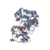
- Assembly
| Deposited unit | 
| ||||||||
|---|---|---|---|---|---|---|---|---|---|
| 1 | 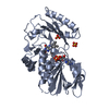
| ||||||||
| 2 | 
| ||||||||
| Unit cell |
|
-Components
| #1: Protein | / Monoamine oxidase-C / MAO-C Mass: 37871.168 Da / Num. of mol.: 2 Source method: isolated from a genetically manipulated source Source: (gene. exp.) Homo sapiens (human) / Gene: RNLS / Plasmid: pET SUMO / Production host:  Escherichia coli (E. coli) Escherichia coli (E. coli)References: UniProt: Q5VYX0, Oxidoreductases; Acting on the CH-NH2 group of donors#2: Chemical | Flavin adenine dinucleotide  Mass: 785.550 Da / Num. of mol.: 2 / Source method: obtained synthetically / Formula: C27H33N9O15P2 / Comment: FAD*YM Mass: 785.550 Da / Num. of mol.: 2 / Source method: obtained synthetically / Formula: C27H33N9O15P2 / Comment: FAD*YM#3: Chemical | Sulfate  Mass: 96.063 Da / Num. of mol.: 3 / Source method: obtained synthetically / Formula: SO4 Mass: 96.063 Da / Num. of mol.: 3 / Source method: obtained synthetically / Formula: SO4#4: Water | ChemComp-HOH / | Water Mass: 18.015 Da / Num. of mol.: 128 / Source method: isolated from a natural source / Formula: H2O Mass: 18.015 Da / Num. of mol.: 128 / Source method: isolated from a natural source / Formula: H2OSequence details | NATURAL VARIANT, SEE UNP DATABASE RNLS_HUMAN | |
|---|
-Experimental details
-Experiment
| Experiment | Method: X-RAY DIFFRACTION / Number of used crystals: 1 |
|---|
- Sample preparation
| Crystal | Density Matthews: 2.86 Å3/Da / Density % sol: 56.92 % |
|---|---|
| Crystal grow | Temperature: 293 K / Method: vapor diffusion, sitting drop / pH: 6.5 Details: 30% PEG 8000, 0.2M ammonium sulfate, pH 6.5, VAPOR DIFFUSION, SITTING DROP, temperature 293K |
-Data collection
| Diffraction | Mean temperature: 100 K |
|---|---|
| Diffraction source | Source: SYNCHROTRON / Site: ESRF  / Beamline: ID23-2 / Wavelength: 0.8726 Å / Beamline: ID23-2 / Wavelength: 0.8726 Å |
| Detector | Type: MARMOSAIC 225 mm CCD / Detector: CCD / Date: Jan 29, 2010 |
| Radiation | Monochromator: horizontally side diffracting Silicon 111 crystal Protocol: SINGLE WAVELENGTH / Monochromatic (M) / Laue (L): M / Scattering type: x-ray |
| Radiation wavelength | Wavelength: 0.8726 Å / Relative weight: 1 |
| Reflection | Resolution: 2.5→48.39 Å / Num. all: 25082 / Num. obs: 29646 / % possible obs: 84.7 % / Observed criterion σ(F): 0 / Observed criterion σ(I): 0 / Biso Wilson estimate: 45.16 Å2 |
| Reflection shell | Resolution: 2.5→2.64 Å / Num. possible: 3770 |
- Processing
| Software |
| ||||||||||||||||||||||||||||||||||||||||||||||||||||||||||||||||||||||||||||||||||||||||||||||||||||||||||||
|---|---|---|---|---|---|---|---|---|---|---|---|---|---|---|---|---|---|---|---|---|---|---|---|---|---|---|---|---|---|---|---|---|---|---|---|---|---|---|---|---|---|---|---|---|---|---|---|---|---|---|---|---|---|---|---|---|---|---|---|---|---|---|---|---|---|---|---|---|---|---|---|---|---|---|---|---|---|---|---|---|---|---|---|---|---|---|---|---|---|---|---|---|---|---|---|---|---|---|---|---|---|---|---|---|---|---|---|---|---|
| Refinement | Method to determine structure: MOLECULAR REPLACEMENT Starting model: 3KKJ Resolution: 2.5→28.94 Å / Cor.coef. Fo:Fc: 0.9218 / Cor.coef. Fo:Fc free: 0.8859 / Occupancy max: 1 / Occupancy min: 0.01 / Cross valid method: THROUGHOUT / σ(F): 0 / Stereochemistry target values: Engh & Huber
| ||||||||||||||||||||||||||||||||||||||||||||||||||||||||||||||||||||||||||||||||||||||||||||||||||||||||||||
| Displacement parameters | Biso max: 136.11 Å2 / Biso mean: 52.77 Å2 / Biso min: 18.02 Å2
| ||||||||||||||||||||||||||||||||||||||||||||||||||||||||||||||||||||||||||||||||||||||||||||||||||||||||||||
| Refine analyze | Luzzati coordinate error obs: 0.369 Å | ||||||||||||||||||||||||||||||||||||||||||||||||||||||||||||||||||||||||||||||||||||||||||||||||||||||||||||
| Refinement step | Cycle: LAST / Resolution: 2.5→28.94 Å
| ||||||||||||||||||||||||||||||||||||||||||||||||||||||||||||||||||||||||||||||||||||||||||||||||||||||||||||
| Refine LS restraints |
| ||||||||||||||||||||||||||||||||||||||||||||||||||||||||||||||||||||||||||||||||||||||||||||||||||||||||||||
| LS refinement shell | Resolution: 2.5→2.6 Å / Total num. of bins used: 13
| ||||||||||||||||||||||||||||||||||||||||||||||||||||||||||||||||||||||||||||||||||||||||||||||||||||||||||||
| Refinement TLS params. | Method: refined / Refine-ID: X-RAY DIFFRACTION
| ||||||||||||||||||||||||||||||||||||||||||||||||||||||||||||||||||||||||||||||||||||||||||||||||||||||||||||
| Refinement TLS group |
|
