 Movie
Movie Controller

[English] 日本語
 Yorodumi
- PDB-3qij: Primitive-monoclinic crystal structure of the FERM domain of prot... -
+ Open data

- Basic information
| Entry | Database: PDB / ID: 3qij | ||||||
|---|---|---|---|---|---|---|---|
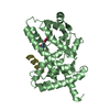
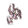
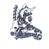
| Title | Primitive-monoclinic crystal structure of the FERM domain of protein 4.1R | ||||||
 Components Components | Protein 4.1 | ||||||
 Keywords Keywords |  STRUCTURAL PROTEIN / cytoskeleton / Structural Genomics / Structural Genomics Consortium / SGC STRUCTURAL PROTEIN / cytoskeleton / Structural Genomics / Structural Genomics Consortium / SGC | ||||||
| Function / homology |  Function and homology information Function and homology information1-phosphatidylinositol binding / regulation of intestinal absorption / spectrin-associated cytoskeleton / actomyosin structure organization / Neurexins and neuroligins / cortical actin cytoskeleton organization / spectrin binding / intercellular bridge / cortical cytoskeleton / regulation of calcium ion transport ...1-phosphatidylinositol binding / regulation of intestinal absorption / spectrin-associated cytoskeleton / actomyosin structure organization / Neurexins and neuroligins / cortical actin cytoskeleton organization / spectrin binding / intercellular bridge / cortical cytoskeleton / regulation of calcium ion transport / positive regulation of protein localization to cell cortex / phosphoprotein binding / structural constituent of cytoskeleton / cytoplasmic side of plasma membrane / mitotic spindle / positive regulation of protein binding / cell junction / actin binding / cell cortex / actin cytoskeleton organization / basolateral plasma membrane / protein-containing complex assembly / cytoskeleton / nuclear body / calmodulin binding / cell cycle / cell division / protein-containing complex / plasma membrane / cytosolSimilarity search - Function | ||||||
| Biological species |  Homo sapiens (human) Homo sapiens (human) | ||||||
| Method | X-RAY DIFFRACTION / SYNCHROTRON / MOLECULAR REPLACEMENT / molecular replacement / Resolution: 1.8 Å | ||||||
 Authors Authors | Nedyalkova, L. / Zhong, N. / Tong, Y. / Tempel, W. / Arrowsmith, C.H. / Edwards, A.M. / Bountra, C. / Weigelt, J. / Park, H. / Structural Genomics Consortium (SGC) | ||||||
 Citation Citation | Journal: to be published Title: Primitive-monoclinic crystal structure of the FERM domain of protein 4.1R Authors: Nedyalkova, L. / Zhong, N. / Tong, Y. / Tempel, W. / Arrowsmith, C.H. / Edwards, A.M. / Bountra, C. / Weigelt, J. / Park, H. | ||||||
| History |
|
- Structure visualization
| Structure viewer | Molecule: MolmilJmol/JSmol |
|---|
- Downloads & links
-Download
| PDBx/mmCIF format | 3qij.cif.gz | 235.4 KB | Display | PDBx/mmCIF format |
|---|---|---|---|---|
| PDB format | pdb3qij.ent.gz | 189.2 KB | Display | PDB format |
| PDBx/mmJSON format | 3qij.json.gz | Tree view | PDBx/mmJSON format | |
| Others |  Other downloads Other downloads |
-Validation report
| Arichive directory | https://data.pdbj.org/pub/pdb/validation_reports/qi/3qijftp://data.pdbj.org/pub/pdb/validation_reports/qi/3qij | HTTPS FTP |
|---|
-Related structure data
| Related structure data | 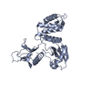 1gg3S S: Starting model for refinement |
|---|---|
| Similar structure data |


-Links
 PDBj
PDBj

- Assembly
| Deposited unit | 
| ||||||||
|---|---|---|---|---|---|---|---|---|---|
| 1 | 
| ||||||||
| 2 | 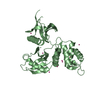
| ||||||||
| Unit cell |
| ||||||||
| Details | AUTHORS STATE THAT THE BIOLOGICAL MOLECULE IS UNKNOWN. |
-Components
| #1: Protein | Mass: 34431.500 Da / Num. of mol.: 2 Source method: isolated from a genetically manipulated source Source: (gene. exp.) Homo sapiens (human) / Gene: EPB41, E41P / Plasmid: pET28-MHL / Production host:  Escherichia coli (E. coli) / Strain (production host): BL21-V2R-pRARE2 / References: UniProt: P11171 Escherichia coli (E. coli) / Strain (production host): BL21-V2R-pRARE2 / References: UniProt: P11171#2: Chemical | ChemComp-UNX /   Num. of mol.: 19 / Source method: obtained synthetically Num. of mol.: 19 / Source method: obtained synthetically#3: Water | ChemComp-HOH / | Water Mass: 18.015 Da / Num. of mol.: 220 / Source method: isolated from a natural source / Formula: H2O Mass: 18.015 Da / Num. of mol.: 220 / Source method: isolated from a natural source / Formula: H2O |
|---|
-Experimental details
-Experiment
| Experiment | Method: X-RAY DIFFRACTION / Number of used crystals: 1 |
|---|
- Sample preparation
| Crystal | Density Matthews: 2.04 Å3/Da / Density % sol: 39.81 % |
|---|---|
| Crystal grow | Temperature: 293 K / Method: vapor diffusion, sitting drop / pH: 7.5 Details: 25% PEG3350, 0.2M magnesium chloride, 1% w/w dispase-I, pH 7.5, vapor diffusion, sitting drop, temperature 293K |
-Data collection
| Diffraction | Mean temperature: 100 K | ||||||||||||||||||||||||||||||||||||||||||||||||||||||||||||||||||||||||||||||||||||||||||||||||||||||||||||||||||||||||||||||
|---|---|---|---|---|---|---|---|---|---|---|---|---|---|---|---|---|---|---|---|---|---|---|---|---|---|---|---|---|---|---|---|---|---|---|---|---|---|---|---|---|---|---|---|---|---|---|---|---|---|---|---|---|---|---|---|---|---|---|---|---|---|---|---|---|---|---|---|---|---|---|---|---|---|---|---|---|---|---|---|---|---|---|---|---|---|---|---|---|---|---|---|---|---|---|---|---|---|---|---|---|---|---|---|---|---|---|---|---|---|---|---|---|---|---|---|---|---|---|---|---|---|---|---|---|---|---|---|
| Diffraction source | Source: SYNCHROTRON / Site: APS  / Beamline: 19-ID / Wavelength: 0.98322 Å / Beamline: 19-ID / Wavelength: 0.98322 Å | ||||||||||||||||||||||||||||||||||||||||||||||||||||||||||||||||||||||||||||||||||||||||||||||||||||||||||||||||||||||||||||||
| Detector | Type: ADSC QUANTUM 315 / Detector: CCD / Date: Nov 27, 2010 | ||||||||||||||||||||||||||||||||||||||||||||||||||||||||||||||||||||||||||||||||||||||||||||||||||||||||||||||||||||||||||||||
| Radiation | Protocol: SINGLE WAVELENGTH / Monochromatic (M) / Laue (L): M / Scattering type: x-ray | ||||||||||||||||||||||||||||||||||||||||||||||||||||||||||||||||||||||||||||||||||||||||||||||||||||||||||||||||||||||||||||||
| Radiation wavelength | Wavelength: 0.98322 Å / Relative weight: 1 | ||||||||||||||||||||||||||||||||||||||||||||||||||||||||||||||||||||||||||||||||||||||||||||||||||||||||||||||||||||||||||||||
| Reflection | Resolution: 1.8→50 Å / Num. obs: 51901 / % possible obs: 99.7 % / Redundancy: 3.6 % / Rmerge(I) obs: 0.055 / Χ2: 1.043 / Net I/σ(I): 10.5 | ||||||||||||||||||||||||||||||||||||||||||||||||||||||||||||||||||||||||||||||||||||||||||||||||||||||||||||||||||||||||||||||
| Reflection shell |
|
-Phasing
| Phasing | Method: molecular replacement |
|---|
- Processing
| Software |
| |||||||||||||||||||||||||||||||||||||||||||||||||||||||||||||||||||||||||||||||||||||||||||||||||||||||||||||||||||||||||||||||||||||||||||||||||||
|---|---|---|---|---|---|---|---|---|---|---|---|---|---|---|---|---|---|---|---|---|---|---|---|---|---|---|---|---|---|---|---|---|---|---|---|---|---|---|---|---|---|---|---|---|---|---|---|---|---|---|---|---|---|---|---|---|---|---|---|---|---|---|---|---|---|---|---|---|---|---|---|---|---|---|---|---|---|---|---|---|---|---|---|---|---|---|---|---|---|---|---|---|---|---|---|---|---|---|---|---|---|---|---|---|---|---|---|---|---|---|---|---|---|---|---|---|---|---|---|---|---|---|---|---|---|---|---|---|---|---|---|---|---|---|---|---|---|---|---|---|---|---|---|---|---|---|---|---|
| Refinement | Method to determine structure: MOLECULAR REPLACEMENT Starting model: pdb entry 1gg3 Resolution: 1.8→30 Å / Cor.coef. Fo:Fc: 0.954 / Cor.coef. Fo:Fc free: 0.919 / WRfactor Rfree: 0.254 / WRfactor Rwork: 0.202 / Occupancy max: 1 / Occupancy min: 0.3 / SU B: 9.195 / SU ML: 0.122 / Cross valid method: THROUGHOUT / σ(F): 0 / ESU R Free: 0.144 / Stereochemistry target values: MAXIMUM LIKELIHOOD Details: HYDROGENS HAVE BEEN ADDED IN THE RIDING POSITIONS. U VALUES WITH TLS ADDED. The programs coot, buccaneer, arp/warp were used during refinement as well as the molprobity server.
| |||||||||||||||||||||||||||||||||||||||||||||||||||||||||||||||||||||||||||||||||||||||||||||||||||||||||||||||||||||||||||||||||||||||||||||||||||
| Solvent computation | Ion probe radii: 0.8 Å / Shrinkage radii: 0.8 Å / VDW probe radii: 1.4 Å / Solvent model: MASK BULK SOLVENT | |||||||||||||||||||||||||||||||||||||||||||||||||||||||||||||||||||||||||||||||||||||||||||||||||||||||||||||||||||||||||||||||||||||||||||||||||||
| Displacement parameters | Biso max: 80.04 Å2 / Biso mean: 25.977 Å2 / Biso min: 13.49 Å2
| |||||||||||||||||||||||||||||||||||||||||||||||||||||||||||||||||||||||||||||||||||||||||||||||||||||||||||||||||||||||||||||||||||||||||||||||||||
| Refinement step | Cycle: LAST / Resolution: 1.8→30 Å
| |||||||||||||||||||||||||||||||||||||||||||||||||||||||||||||||||||||||||||||||||||||||||||||||||||||||||||||||||||||||||||||||||||||||||||||||||||
| Refine LS restraints |
| |||||||||||||||||||||||||||||||||||||||||||||||||||||||||||||||||||||||||||||||||||||||||||||||||||||||||||||||||||||||||||||||||||||||||||||||||||
| LS refinement shell | Refine-ID: X-RAY DIFFRACTION / Total num. of bins used: 20
| |||||||||||||||||||||||||||||||||||||||||||||||||||||||||||||||||||||||||||||||||||||||||||||||||||||||||||||||||||||||||||||||||||||||||||||||||||
| Refinement TLS params. | Method: refined / Refine-ID: X-RAY DIFFRACTION
| |||||||||||||||||||||||||||||||||||||||||||||||||||||||||||||||||||||||||||||||||||||||||||||||||||||||||||||||||||||||||||||||||||||||||||||||||||
| Refinement TLS group |
|
