 Movie
Movie Controller


+ Open data

- Basic information
| Entry | Database: PDB / ID: 3qic | ||||||
|---|---|---|---|---|---|---|---|
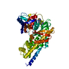
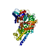
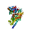
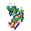
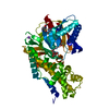
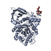
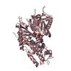
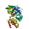
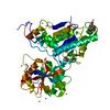
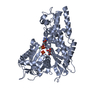
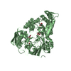
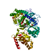
| Title | The structure of human glucokinase E339K mutation | ||||||
 Components Components | Glucokinase | ||||||
 Keywords Keywords | TRANSFERASE / Glycolysis / Kinase / Sugar Binding / Phosphorylation | ||||||
| Function / homology |  Function and homology information Function and homology informationDefective GCK causes maturity-onset diabetes of the young 2 (MODY2) / mannokinase activity / glucose sensor activity / regulation of potassium ion transport / hexokinase / fructokinase activity / carbohydrate phosphorylation / glucokinase activity / glucose catabolic process / glucose 6-phosphate metabolic process ...Defective GCK causes maturity-onset diabetes of the young 2 (MODY2) / mannokinase activity / glucose sensor activity / regulation of potassium ion transport / hexokinase / fructokinase activity / carbohydrate phosphorylation / glucokinase activity / glucose catabolic process / glucose 6-phosphate metabolic process / NADP metabolic process / Regulation of Glucokinase by Glucokinase Regulatory Protein / Defective TPR may confer susceptibility towards thyroid papillary carcinoma (TPC) / D-glucose binding / cellular response to leptin stimulus / calcium ion import / canonical glycolysis / Glycolysis / regulation of glycolytic process / intracellular glucose homeostasis / Regulation of gene expression in beta cells / regulation of insulin secretion / positive regulation of glycogen biosynthetic process / FOXO-mediated transcription of oxidative stress, metabolic and neuronal genes / negative regulation of gluconeogenesis / response to glucose / glycolytic process / positive regulation of insulin secretion / cellular response to insulin stimulus / glucose metabolic process / glucose homeostasis / mitochondrion / nucleoplasm / ATP binding / cytosolSimilarity search - Function | ||||||
| Biological species |  Homo sapiens (human) Homo sapiens (human) | ||||||
| Method | X-RAY DIFFRACTION / SYNCHROTRON / MOLECULAR REPLACEMENT / Resolution: 2.2 Å | ||||||
 Authors Authors | Liu, Q. / Liu, S. / Liu, J. | ||||||
 Citation Citation | Journal: Febs Lett. / Year: 2011 Title: Crystal structure of E339K mutated human glucokinase reveals changes in the ATP binding site. Authors: Liu, Q. / Shen, Y. / Liu, S. / Weng, J. / Liu, J. | ||||||
| History |
|
- Structure visualization
| Structure viewer | Molecule: MolmilJmol/JSmol |
|---|
- Downloads & links
-Download
| PDBx/mmCIF format | 3qic.cif.gz | 104.1 KB | Display | PDBx/mmCIF format |
|---|---|---|---|---|
| PDB format | pdb3qic.ent.gz | 79.1 KB | Display | PDB format |
| PDBx/mmJSON format | 3qic.json.gz | Tree view | PDBx/mmJSON format | |
| Others |  Other downloads Other downloads |
-Validation report
| Arichive directory | https://data.pdbj.org/pub/pdb/validation_reports/qi/3qicftp://data.pdbj.org/pub/pdb/validation_reports/qi/3qic | HTTPS FTP |
|---|
-Related structure data
| Related structure data | 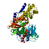 1v4sS S: Starting model for refinement |
|---|---|
| Similar structure data |
-Links
 PDBj
PDBj


- Assembly
| Deposited unit | 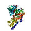
| ||||||||
|---|---|---|---|---|---|---|---|---|---|
| 1 |
| ||||||||
| Unit cell |
|
-Components
| #1: Protein | / Hexokinase type IV / HK IV / Hexokinase-4 / HK4 / Hexokinase-D Mass: 53076.230 Da / Num. of mol.: 1 / Fragment: residues 12-465 / Mutation: E339K Source method: isolated from a genetically manipulated source Source: (gene. exp.) Homo sapiens (human) / Gene: GCK / Plasmid: pET21a / Production host:  Escherichia coli (E. coli) / Strain (production host): Bl21 / References: UniProt: P35557, glucokinase Escherichia coli (E. coli) / Strain (production host): Bl21 / References: UniProt: P35557, glucokinase | ||
|---|---|---|---|
| #2: Sugar | ChemComp-GLC / Glucose  Type: D-saccharide, alpha linking / Mass: 180.156 Da / Num. of mol.: 1 Type: D-saccharide, alpha linking / Mass: 180.156 Da / Num. of mol.: 1Source method: isolated from a genetically manipulated source Formula: C6H12O6 | ||
| #3: Chemical | ChemComp-GOL / Glycerol  Mass: 92.094 Da / Num. of mol.: 6 / Source method: obtained synthetically / Formula: C3H8O3 Mass: 92.094 Da / Num. of mol.: 6 / Source method: obtained synthetically / Formula: C3H8O3#4: Water | ChemComp-HOH / | Water Mass: 18.015 Da / Num. of mol.: 51 / Source method: isolated from a natural source / Formula: H2O Mass: 18.015 Da / Num. of mol.: 51 / Source method: isolated from a natural source / Formula: H2O |
-Experimental details
-Experiment
| Experiment | Method: X-RAY DIFFRACTION / Number of used crystals: 1 |
|---|
- Sample preparation
| Crystal | Density Matthews: 2.74 Å3/Da / Density % sol: 55.09 % |
|---|---|
| Crystal grow | Temperature: 293 K / Method: vapor diffusion, sitting drop / pH: 7.3 Details: 25% (w/v) PEG3350, 0.1M HEPES pH 7.3, 0.2M ammonium sulfate, VAPOR DIFFUSION, SITTING DROP, temperature 293K |
-Data collection
| Diffraction | Mean temperature: 100 K | |||||||||||||||||||||
|---|---|---|---|---|---|---|---|---|---|---|---|---|---|---|---|---|---|---|---|---|---|---|
| Diffraction source | Source: SYNCHROTRON / Site: SSRF  / Beamline: BL17U / Beamline: BL17U | |||||||||||||||||||||
| Detector | Type: RAYONIX MX-225 / Detector: CCD | |||||||||||||||||||||
| Radiation | Protocol: SINGLE WAVELENGTH / Monochromatic (M) / Laue (L): M / Scattering type: x-ray | |||||||||||||||||||||
| Radiation wavelength | Relative weight: 1 | |||||||||||||||||||||
| Reflection | Resolution: 2.2→41.19 Å / Num. obs: 30831 / % possible obs: 99.9 % / Observed criterion σ(I): 1 / Redundancy: 6 % / Biso Wilson estimate: 40.9 Å2 / Rmerge(I) obs: 0.077 | |||||||||||||||||||||
| Reflection shell |
|
- Processing
| Software |
| ||||||||||||||||||||||||||||||||||||||||||||||||||||||||||||||||||
|---|---|---|---|---|---|---|---|---|---|---|---|---|---|---|---|---|---|---|---|---|---|---|---|---|---|---|---|---|---|---|---|---|---|---|---|---|---|---|---|---|---|---|---|---|---|---|---|---|---|---|---|---|---|---|---|---|---|---|---|---|---|---|---|---|---|---|---|
| Refinement | Method to determine structure: MOLECULAR REPLACEMENT Starting model: 1V4S Resolution: 2.2→39.375 Å / SU ML: 0.29 / σ(F): 0 / Stereochemistry target values: ML
| ||||||||||||||||||||||||||||||||||||||||||||||||||||||||||||||||||
| Solvent computation | Shrinkage radii: 0.95 Å / VDW probe radii: 1.2 Å / Solvent model: FLAT BULK SOLVENT MODEL / Bsol: 47.626 Å2 / ksol: 0.385 e/Å3 | ||||||||||||||||||||||||||||||||||||||||||||||||||||||||||||||||||
| Displacement parameters |
| ||||||||||||||||||||||||||||||||||||||||||||||||||||||||||||||||||
| Refinement step | Cycle: LAST / Resolution: 2.2→39.375 Å
| ||||||||||||||||||||||||||||||||||||||||||||||||||||||||||||||||||
| Refine LS restraints |
| ||||||||||||||||||||||||||||||||||||||||||||||||||||||||||||||||||
| LS refinement shell | Refine-ID: X-RAY DIFFRACTION / Total num. of bins used: 10
|
