 Movie
Movie Controller

[English] 日本語
 Yorodumi
- PDB-3qhm: Crystal analysis of the complex structure, E342A-cellotetraose, o... -
+ Open data

- Basic information
| Entry | Database: PDB / ID: 3qhm | |||||||||
|---|---|---|---|---|---|---|---|---|---|---|
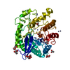
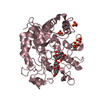
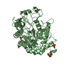
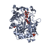
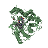
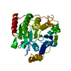
| Title | Crystal analysis of the complex structure, E342A-cellotetraose, of endocellulase from pyrococcus horikoshii | |||||||||
 Components Components | 458aa long hypothetical endo-1,4-beta-glucanase | |||||||||
 Keywords Keywords |  HYDROLASE / cellulase / endoglucanase / catalytic domain HYDROLASE / cellulase / endoglucanase / catalytic domain | |||||||||
| Function / homology |  Function and homology information Function and homology informationhydrolase activity, hydrolyzing O-glycosyl compounds / carbohydrate metabolic process / metal ion bindingSimilarity search - Function | |||||||||
| Biological species |   Pyrococcus horikoshii (archaea) Pyrococcus horikoshii (archaea) | |||||||||
| Method | X-RAY DIFFRACTION / SYNCHROTRON / MOLECULAR REPLACEMENT / Resolution: 2.01 Å | |||||||||
 Authors Authors | Kim, H.-W. / Ishikawa, K. | |||||||||
 Citation Citation | Journal: Biochem.J. / Year: 2011 Title: Functional analysis of hyperthermophilic endocellulase from Pyrococcus horikoshii by crystallographic snapshots Authors: Kim, H.-W. / Ishikawa, K. #1: Journal: Acta Crystallogr.,Sect.F / Year: 2008 Title: Crystallization and preliminary X-ray analysis of endoglucanase from Pyrococcus horikoshii Authors: Kim, H.-W. / Mino, K. / Ishikawa, K. #2: Journal: Proteins / Year: 2010Title: Structure of hyperthermophilic endocellulase from Pyrococcus horikoshii Authors: Kim, H.-W. / Ishikawa, K. | |||||||||
| History |
|
- Structure visualization
| Structure viewer | Molecule: MolmilJmol/JSmol |
|---|
- Downloads & links
-Download
| PDBx/mmCIF format | 3qhm.cif.gz | 244.7 KB | Display | PDBx/mmCIF format |
|---|---|---|---|---|
| PDB format | pdb3qhm.ent.gz | 197.8 KB | Display | PDB format |
| PDBx/mmJSON format | 3qhm.json.gz | Tree view | PDBx/mmJSON format | |
| Others |  Other downloads Other downloads |
-Validation report
| Arichive directory | https://data.pdbj.org/pub/pdb/validation_reports/qh/3qhmftp://data.pdbj.org/pub/pdb/validation_reports/qh/3qhm | HTTPS FTP |
|---|
-Related structure data
| Related structure data |  3axxC  3qhnC  3qhoC  2zumS C: citing same article ( S: Starting model for refinement |
|---|---|
| Similar structure data |


-Links
 PDBj
PDBj- Assembly
| Deposited unit | 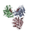
| ||||||||
|---|---|---|---|---|---|---|---|---|---|
| 1 | 
| ||||||||
| 2 | 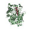
| ||||||||
| 3 | 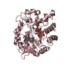
| ||||||||
| Unit cell |
|
-Components
| #1: Protein | Mass: 51924.844 Da / Num. of mol.: 3 / Mutation: E342A Source method: isolated from a genetically manipulated source Source: (gene. exp.) Pyrococcus horikoshii (archaea) / Strain: OT3 / Gene: PH1171 / Plasmid: PET11A / Production host:  ESCHERICHIA COLI (E. coli) / Strain (production host): BL21(DE3) / References: UniProt: O58925, cellulase ESCHERICHIA COLI (E. coli) / Strain (production host): BL21(DE3) / References: UniProt: O58925, cellulase#2: Polysaccharide | 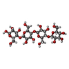  , Oligosaccharide / Class: Metabolism / Mass: 666.578 Da / Num. of mol.: 3 , Oligosaccharide / Class: Metabolism / Mass: 666.578 Da / Num. of mol.: 3Source method: isolated from a genetically manipulated source Details: oligosaccharide / References: beta-cellotetraose #3: Polysaccharide | beta-D-glucopyranose-(1-4)-beta-D-glucopyranose / beta-cellobiose |   , Oligosaccharide / Class: Metabolism / Mass: 342.297 Da / Num. of mol.: 1 , Oligosaccharide / Class: Metabolism / Mass: 342.297 Da / Num. of mol.: 1Source method: isolated from a genetically manipulated source Details: oligosaccharide / References: beta-cellobiose #4: Water | ChemComp-HOH / | Water Mass: 18.015 Da / Num. of mol.: 206 / Source method: isolated from a natural source / Formula: H2O Mass: 18.015 Da / Num. of mol.: 206 / Source method: isolated from a natural source / Formula: H2O |
|---|
-Experimental details
-Experiment
| Experiment | Method: X-RAY DIFFRACTION / Number of used crystals: 1 |
|---|
- Sample preparation
| Crystal | Density Matthews: 2 Å3/Da / Density % sol: 38.57 % |
|---|---|
| Crystal grow | Temperature: 295 K / Method: vapor diffusion, hanging drop / pH: 6.5 Details: 1.5M AMMONIUM PHOSPHATE, 0.1M MES BUFFER, pH 6.5, VAPOR DIFFUSION, HANGING DROP, temperature 295K |
-Data collection
| Diffraction | Mean temperature: 100 K |
|---|---|
| Diffraction source | Source: SYNCHROTRON / Site: SPring-8  / Beamline: BL38B1 / Wavelength: 1 / Beamline: BL38B1 / Wavelength: 1 |
| Detector | Type: RIGAKU JUPITER 210 / Detector: CCD / Date: Jul 3, 2008 |
| Radiation | Protocol: SINGLE WAVELENGTH / Monochromatic (M) / Laue (L): M / Scattering type: x-ray |
| Radiation wavelength | Wavelength: 1 Å / Relative weight: 1 |
| Reflection | Resolution: 2→50 Å / Num. obs: 73075 / % possible obs: 88.3 % / Redundancy: 2.9 % / Biso Wilson estimate: 16.1 Å2 / Rmerge(I) obs: 0.066 / Net I/σ(I): 17.6 |
| Reflection shell | Resolution: 2→2.03 Å / Redundancy: 2.2 % / Rmerge(I) obs: 0.328 / % possible all: 77.8 |
- Processing
| Software |
| ||||||||||||||||||||||||||||||||||||||||||||||||||||||||||||
|---|---|---|---|---|---|---|---|---|---|---|---|---|---|---|---|---|---|---|---|---|---|---|---|---|---|---|---|---|---|---|---|---|---|---|---|---|---|---|---|---|---|---|---|---|---|---|---|---|---|---|---|---|---|---|---|---|---|---|---|---|---|
| Refinement | Method to determine structure: MOLECULAR REPLACEMENT Starting model: PDB ENTRY 2ZUM Resolution: 2.01→35.25 Å / Rfactor Rfree error: 0.004 / Data cutoff high absF: 537235.61 / Data cutoff low absF: 0 / Isotropic thermal model: RESTRAINED / Cross valid method: THROUGHOUT / σ(F): 0 / Details: BULK SOLVENT MODEL USED
| ||||||||||||||||||||||||||||||||||||||||||||||||||||||||||||
| Solvent computation | Solvent model: FLAT MODEL / Bsol: 70.18 Å2 / ksol: 0.4 e/Å3 | ||||||||||||||||||||||||||||||||||||||||||||||||||||||||||||
| Displacement parameters | Biso mean: 48.5 Å2
| ||||||||||||||||||||||||||||||||||||||||||||||||||||||||||||
| Refine analyze |
| ||||||||||||||||||||||||||||||||||||||||||||||||||||||||||||
| Refinement step | Cycle: LAST / Resolution: 2.01→35.25 Å
| ||||||||||||||||||||||||||||||||||||||||||||||||||||||||||||
| Refine LS restraints |
| ||||||||||||||||||||||||||||||||||||||||||||||||||||||||||||
| LS refinement shell | Resolution: 2.01→2.14 Å / Rfactor Rfree error: 0.018 / Total num. of bins used: 6
| ||||||||||||||||||||||||||||||||||||||||||||||||||||||||||||
| Xplor file |
|
