[English] 日本語
Yorodumi
- PDB-3qc9: Crystal structure of cross-linked bovine GRK1 T8C/N480C double mu... -

+
Open data


ID or keywords:

Loading...

-
Basic information

Entry
Database: PDB / ID: 3qc9
Title
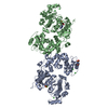
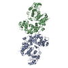
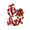
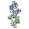
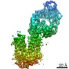
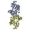
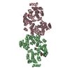
Crystal structure of cross-linked bovine GRK1 T8C/N480C double mutant complexed with ADP and Mg
ComponentsRhodopsin kinase
KeywordsTRANSFERASE / enkaryotic protein kinase fold / protein serine/threonine kinase
Function / homology
Function and homology information


rhodopsin kinase / rhodopsin kinase activity / regulation of opsin-mediated signaling pathway / Inactivation, recovery and regulation of the phototransduction cascade / regulation of signal transduction / visual perception / photoreceptor disc membrane / protein autophosphorylation / signal transduction / ATP binding / cytoplasm
Similarity search - Function
Rhodopsin kinase, catalytic domain / GPCR kinase / Regulator of G-protein Signalling 4, domain 2 / Regulator of G-protein Signalling 4; domain 2 / Regulator of G protein signaling domain / RGS, subdomain 2 / RGS domain / RGS domain profile. / Regulator of G protein signalling domain / RGS domain superfamily ...Rhodopsin kinase, catalytic domain / GPCR kinase / Regulator of G-protein Signalling 4, domain 2 / Regulator of G-protein Signalling 4; domain 2 / Regulator of G protein signaling domain / RGS, subdomain 2 / RGS domain / RGS domain profile. / Regulator of G protein signalling domain / RGS domain superfamily / Extension to Ser/Thr-type protein kinases / AGC-kinase, C-terminal / AGC-kinase C-terminal domain profile. / Transferase(Phosphotransferase) domain 1 / Transferase(Phosphotransferase); domain 1 / Phosphorylase Kinase; domain 1 / Phosphorylase Kinase; domain 1 / Serine/threonine-protein kinase, active site / Serine/Threonine protein kinases active-site signature. / Protein kinase domain / Serine/Threonine protein kinases, catalytic domain / Protein kinase, ATP binding site / Protein kinases ATP-binding region signature. / Protein kinase domain profile. / Protein kinase domain / Protein kinase-like domain superfamily / 2-Layer Sandwich / Orthogonal Bundle / Mainly Alpha / Alpha Beta
Similarity search - Domain/homology
ADENOSINE-5'-DIPHOSPHATE / Rhodopsin kinase GRK1
Similarity search - Component
Biological speciesBos taurus (cattle)
MethodX-RAY DIFFRACTION / SYNCHROTRON / MOLECULAR REPLACEMENT / Resolution: 2.7 Å
AuthorsHuang, C.-C. / Tesmer, J.J.G.
CitationJournal: Biochemistry / Year: 2011
Title: Activation of G Protein-Coupled Receptor Kinase 1 Involves Interactions between Its N-Terminal Region and Its Kinase Domain.
Authors: Huang, C.C. / Orban, T. / Jastrzebska, B. / Palczewski, K. / Tesmer, J.J.
History
DepositionJan 15, 2011Deposition site: RCSB / Processing site: RCSB
Revision 1.0Feb 16, 2011Provider: repository / Type: Initial release
Revision 1.1Jul 13, 2011Group: Version format compliance
Revision 1.2Sep 13, 2023Group: Data collection / Database references ...Data collection / Database references / Derived calculations / Refinement description
Category: chem_comp_atom / chem_comp_bond ...chem_comp_atom / chem_comp_bond / database_2 / pdbx_initial_refinement_model / pdbx_struct_conn_angle / struct_conn / struct_ref_seq_dif / struct_site
Item: _database_2.pdbx_DOI / _database_2.pdbx_database_accession ..._database_2.pdbx_DOI / _database_2.pdbx_database_accession / _pdbx_struct_conn_angle.ptnr1_auth_asym_id / _pdbx_struct_conn_angle.ptnr1_auth_comp_id / _pdbx_struct_conn_angle.ptnr1_auth_seq_id / _pdbx_struct_conn_angle.ptnr1_label_asym_id / _pdbx_struct_conn_angle.ptnr1_label_atom_id / _pdbx_struct_conn_angle.ptnr1_label_comp_id / _pdbx_struct_conn_angle.ptnr1_label_seq_id / _pdbx_struct_conn_angle.ptnr2_auth_asym_id / _pdbx_struct_conn_angle.ptnr2_auth_seq_id / _pdbx_struct_conn_angle.ptnr2_label_asym_id / _pdbx_struct_conn_angle.ptnr3_auth_asym_id / _pdbx_struct_conn_angle.ptnr3_auth_comp_id / _pdbx_struct_conn_angle.ptnr3_auth_seq_id / _pdbx_struct_conn_angle.ptnr3_label_asym_id / _pdbx_struct_conn_angle.ptnr3_label_atom_id / _pdbx_struct_conn_angle.ptnr3_label_comp_id / _pdbx_struct_conn_angle.ptnr3_label_seq_id / _pdbx_struct_conn_angle.value / _struct_conn.pdbx_dist_value / _struct_conn.ptnr1_auth_asym_id / _struct_conn.ptnr1_auth_comp_id / _struct_conn.ptnr1_auth_seq_id / _struct_conn.ptnr1_label_asym_id / _struct_conn.ptnr1_label_atom_id / _struct_conn.ptnr1_label_comp_id / _struct_conn.ptnr1_label_seq_id / _struct_conn.ptnr2_auth_asym_id / _struct_conn.ptnr2_auth_comp_id / _struct_conn.ptnr2_auth_seq_id / _struct_conn.ptnr2_label_asym_id / _struct_conn.ptnr2_label_atom_id / _struct_conn.ptnr2_label_comp_id / _struct_ref_seq_dif.details / _struct_site.pdbx_auth_asym_id / _struct_site.pdbx_auth_comp_id / _struct_site.pdbx_auth_seq_id

-
Structure visualization

Structure viewerMolecule:
MolmilJmol/JSmol

Downloads & links

-
Assembly

Deposited unit
A: Rhodopsin kinase
B: Rhodopsin kinase
C: Rhodopsin kinase
D: Rhodopsin kinase
hetero molecules


Theoretical massNumber of molelcules
Total (without water)247,77118
Polymers245,6844
Non-polymers2,08714
Water2,036113
1
A: Rhodopsin kinase
D: Rhodopsin kinase
hetero molecules


Theoretical massNumber of molelcules
Total (without water)123,8869
Polymers122,8422
Non-polymers1,0447
Water362
TypeNameSymmetry operationNumber
identity operation1_555x,y,z1
Buried area4450 Å2
ΔGint-54 kcal/mol
Surface area45290 Å2
MethodPISA
2
B: Rhodopsin kinase
C: Rhodopsin kinase
hetero molecules


Theoretical massNumber of molelcules
Total (without water)123,8869
Polymers122,8422
Non-polymers1,0447
Water362
TypeNameSymmetry operationNumber
identity operation1_555x,y,z1
Buried area4450 Å2
ΔGint-54 kcal/mol
Surface area45060 Å2
MethodPISA
Unit cell
Length a, b, c (Å)58.696, 89.965, 122.638
Angle α, β, γ (deg.)88.07, 90.26, 68.78
Int Tables number1
Space group name H-MP1
Noncrystallographic symmetry (NCS)NCS domain:
IDEns-ID
11
21
31
41

NCS domain segments:
Dom-IDComponent-IDEns-IDSelection details
111chain A and resid 31:533
211chain B and resid 31:533
311chain C and resid 31:533
411chain D and resid 31:533
DetailsACCORDING TO THE AUTHORS THE BIOLOGICAL ASSEMBLY IS UNKNOWN. HOWEVER, THE MONOMERIC STATE IS FUNCTIONAL IN SOLUTION

-
Components

#1: Protein
Rhodopsin kinase / / RK / G protein-coupled receptor kinase 1


Mass: 61421.012 Da / Num. of mol.: 4 / Fragment: UNP residues 1-535 / Mutation: T8C, N480C
Source method: isolated from a genetically manipulated source
Source: (gene. exp.) Bos taurus (cattle) / Gene: GRK1, RHOK / Cell line (production host): High 5 / Production host: Spodoptera frugiperda (fall armyworm) / References: UniProt: P28327, rhodopsin kinase
#2: Chemical
ChemComp-ADP / ADENOSINE-5'-DIPHOSPHATE / Adenosine diphosphate


Mass: 427.201 Da / Num. of mol.: 4 / Source method: obtained synthetically / Formula: C10H15N5O10P2 / Comment: ADP, energy-carrying molecule*YM
#3: Chemical
ChemComp-MG / MAGNESIUM ION


Mass: 24.305 Da / Num. of mol.: 8 / Source method: obtained synthetically / Formula: Mg
#4: Chemical ChemComp-GOL / GLYCEROL / GLYCERIN / PROPANE-1,2,3-TRIOL / Glycerol


Mass: 92.094 Da / Num. of mol.: 2 / Source method: obtained synthetically / Formula: C3H8O3
#5: Water ChemComp-HOH / water / Water


Mass: 18.015 Da / Num. of mol.: 113 / Source method: isolated from a natural source / Formula: H2O

-
Experimental details

-
Experiment

ExperimentMethod: X-RAY DIFFRACTION / Number of used crystals: 1

-
Sample preparation

CrystalDensity Matthews: 2.46 Å3/Da / Density % sol: 49.91 %
Crystal growTemperature: 277 K / Method: vapor diffusion, hanging drop / pH: 5.5
Details: 14-19% PEG 3350, 0.1 M Bis-Tris pH 5.5, 0.15 M MgCl2, and 0.02% n-dodecyl- D-maltoside, VAPOR DIFFUSION, HANGING DROP, temperature 277K

-
Data collection

DiffractionMean temperature: 100 K
Diffraction sourceSource: SYNCHROTRON / Site: APS / Beamline: 21-ID-G / Wavelength: 0.97856 Å
DetectorType: MARMOSAIC 300 mm CCD / Detector: CCD / Date: Nov 2, 2009
RadiationMonochromator: diamond laue monochromator / Protocol: SINGLE WAVELENGTH / Monochromatic (M) / Laue (L): M / Scattering type: x-ray
Radiation wavelengthWavelength: 0.97856 Å / Relative weight: 1
ReflectionResolution: 2.7→30 Å / Num. all: 64625 / Num. obs: 61717 / % possible obs: 95.5 % / Observed criterion σ(F): 0 / Observed criterion σ(I): -3 / Redundancy: 1.9 % / Rmerge(I) obs: 0.064 / Rsym value: 0.064 / Net I/σ(I): 15
Reflection shellResolution: 2.7→2.75 Å / Redundancy: 1.9 % / Rmerge(I) obs: 0.451 / Mean I/σ(I) obs: 1.6 / Rsym value: 0.451 / % possible all: 95.8

-
Processing

Software
NameVersionClassification
HKL-2000data collection
PHASERphasing
PHENIX(phenix.refine: 1.6.4_486)refinement
HKL-2000data reduction
HKL-2000data scaling
RefinementMethod to determine structure: MOLECULAR REPLACEMENT
Starting model: PDB entry 3C4Z
Resolution: 2.7→29.599 Å / SU ML: 0.34 / σ(F): 1 / Phase error: 26 / Stereochemistry target values: ML
RfactorNum. reflection% reflection
Rfree0.2286 2897 5.06 %
Rwork0.1924 --
obs0.1943 57246 89.18 %
all-64191 -
Solvent computationShrinkage radii: 0.83 Å / VDW probe radii: 1.1 Å / Solvent model: FLAT BULK SOLVENT MODEL / Bsol: 39.025 Å2 / ksol: 0.325 e/Å3
Displacement parameters
Baniso -1Baniso -2Baniso -3
1-11.1488 Å26.3631 Å2-1.1873 Å2
2--8.4486 Å23.6521 Å2
3----19.5974 Å2
Refinement stepCycle: LAST / Resolution: 2.7→29.599 Å
ProteinNucleic acidLigandSolventTotal
Num. atoms15565 0 128 113 15806
Refine LS restraints
Refine-IDTypeDev idealNumber
X-RAY DIFFRACTIONf_bond_d0.00816094
X-RAY DIFFRACTIONf_angle_d1.19821727
X-RAY DIFFRACTIONf_dihedral_angle_d17.1046055
X-RAY DIFFRACTIONf_chiral_restr0.0752269
X-RAY DIFFRACTIONf_plane_restr0.0042825
Refine LS restraints NCS
Ens-IDDom-IDAuth asym-IDNumberRefine-IDTypeRms dev position (Å)
11A3873X-RAY DIFFRACTIONPOSITIONAL
12B3873X-RAY DIFFRACTIONPOSITIONAL0.043
13C3880X-RAY DIFFRACTIONPOSITIONAL0.036
14D3905X-RAY DIFFRACTIONPOSITIONAL0.044
LS refinement shell
Resolution (Å)Rfactor RfreeNum. reflection RfreeRfactor RworkNum. reflection RworkRefine-ID% reflection obs (%)
2.7-2.79650.3882470.30854726X-RAY DIFFRACTION77
2.7965-2.90830.33432780.28034926X-RAY DIFFRACTION81
2.9083-3.04060.34572730.25595102X-RAY DIFFRACTION84
3.0406-3.20070.2672600.2395288X-RAY DIFFRACTION87
3.2007-3.4010.28363070.21365395X-RAY DIFFRACTION88
3.401-3.66310.25123080.20065533X-RAY DIFFRACTION91
3.6631-4.03090.24553040.18815632X-RAY DIFFRACTION93
4.0309-4.61230.17853030.15375826X-RAY DIFFRACTION95
4.6123-5.80380.18433090.15825963X-RAY DIFFRACTION98
5.8038-29.60070.17713080.17545958X-RAY DIFFRACTION98
Refinement TLS params.

Method: refined / Refine-ID: X-RAY DIFFRACTION

IDL112)L122)L132)L222)L232)L332)S11 (Å °)S12 (Å °)S13 (Å °)S21 (Å °)S22 (Å °)S23 (Å °)S31 (Å °)S32 (Å °)S33 (Å °)T112)T122)T132)T222)T232)T332)Origin x (Å)Origin y (Å)Origin z (Å)
10.1174-0.02090.17930.0677-0.10871.10050.06260.0452-0.0234-0.0257-0.00050.03730.1710.1813-0.06240.22380.041-0.0270.2234-0.02610.2107-30.2211-10.753649.6961
20.2133-0.0530.3562-0.10.05891.26730.00620.02360.0021-0.00870.03370.00660.0770.0776-0.03270.2546-0.0380.00740.2139-0.00040.2447-42.1369-53.044844.4895
30.04690.01920.11160.0697-0.16250.46760.0317-0.0124-0.00190.02460.0093-0.0252-0.0824-0.033-0.03590.22140.03030.00840.19750.01930.2321-53.9075-51.7895-13.2996
40.2234-0.1104-0.1365-0.00610.25010.8380.02450.02870.0163-0.00780.0029-0.0219-0.15820.072300.2003-0.02030.0080.16870.01350.2035-41.6764-9.504-15.4474
Refinement TLS group
IDRefine-IDRefine TLS-IDSelection details
1X-RAY DIFFRACTION1chain A
2X-RAY DIFFRACTION2chain B
3X-RAY DIFFRACTION3chain C
4X-RAY DIFFRACTION4chain D

+
About Yorodumi

-
News

-
Feb 9, 2022. New format data for meta-information of EMDB entries

New format data for meta-information of EMDB entries

  • Version 3 of the EMDB header file is now the official format.
  • The previous official version 1.9 will be removed from the archive.

Related info.:EMDB header

External links:wwPDB to switch to version 3 of the EMDB data model

-
Aug 12, 2020. Covid-19 info

Covid-19 info

URL: https://pdbj.org/emnavi/covid19.php

New page: Covid-19 featured information page in EM Navigator.

Related info.:Covid-19 info / Mar 5, 2020. Novel coronavirus structure data

+
Mar 5, 2020. Novel coronavirus structure data

Novel coronavirus structure data

Related info.:Yorodumi Speices / Aug 12, 2020. Covid-19 info

External links:COVID-19 featured content - PDBj / Molecule of the Month (242):Coronavirus Proteases

+
Jan 31, 2019. EMDB accession codes are about to change! (news from PDBe EMDB page)

EMDB accession codes are about to change! (news from PDBe EMDB page)

  • The allocation of 4 digits for EMDB accession codes will soon come to an end. Whilst these codes will remain in use, new EMDB accession codes will include an additional digit and will expand incrementally as the available range of codes is exhausted. The current 4-digit format prefixed with “EMD-” (i.e. EMD-XXXX) will advance to a 5-digit format (i.e. EMD-XXXXX), and so on. It is currently estimated that the 4-digit codes will be depleted around Spring 2019, at which point the 5-digit format will come into force.
  • The EM Navigator/Yorodumi systems omit the EMD- prefix.

Related info.:Q: What is EMD? / ID/Accession-code notation in Yorodumi/EM Navigator

External links:EMDB Accession Codes are Changing Soon! / Contact to PDBj

+
Jul 12, 2017. Major update of PDB

Major update of PDB

  • wwPDB released updated PDB data conforming to the new PDBx/mmCIF dictionary.
  • This is a major update changing the version number from 4 to 5, and with Remediation, in which all the entries are updated.
  • In this update, many items about electron microscopy experimental information are reorganized (e.g. em_software).
  • Now, EM Navigator and Yorodumi are based on the updated data.

External links:wwPDB Remediation / Enriched Model Files Conforming to OneDep Data Standards Now Available in the PDB FTP Archive

-
Yorodumi

Thousand views of thousand structures

  • Yorodumi is a browser for structure data from EMDB, PDB, SASBDB, etc.
  • This page is also the successor to EM Navigator detail page, and also detail information page/front-end page for Omokage search.
  • The word "yorodu" (or yorozu) is an old Japanese word meaning "ten thousand". "mi" (miru) is to see.

Related info.:EMDB / PDB / SASBDB / Comparison of 3 databanks / Yorodumi Search / Aug 31, 2016. New EM Navigator & Yorodumi / Yorodumi Papers / Jmol/JSmol / Function and homology information / Changes in new EM Navigator and Yorodumi

Read more