 Movie
Movie Controller


+ Open data

- Basic information
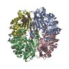
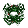
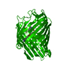
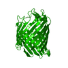
| Entry | Database: PDB / ID: 3q98 | ||||||
|---|---|---|---|---|---|---|---|
| Title | Structure of ygeW encoded protein from E. coli | ||||||
 Components Components | transcarbamylase | ||||||
 Keywords Keywords |  TRANSFERASE / Rossmann Fold / Unknown transcarbamylase TRANSFERASE / Rossmann Fold / Unknown transcarbamylase | ||||||
| Function / homology |  Function and homology informationTransferases; Transferring one-carbon groups; Carboxy- and carbamoyltransferases / carboxyl- or carbamoyltransferase activity / ornithine carbamoyltransferase activity / arginine biosynthetic process via ornithine / citrulline biosynthetic process / amino acid metabolic process / amino acid binding Function and homology informationTransferases; Transferring one-carbon groups; Carboxy- and carbamoyltransferases / carboxyl- or carbamoyltransferase activity / ornithine carbamoyltransferase activity / arginine biosynthetic process via ornithine / citrulline biosynthetic process / amino acid metabolic process / amino acid bindingSimilarity search - Function | ||||||
| Biological species |  Escherichia coli (E. coli) Escherichia coli (E. coli) | ||||||
| Method | X-RAY DIFFRACTION / SYNCHROTRON / SAD / Resolution: 2.001 Å | ||||||
 Authors Authors | Li, Y. / Jing, Z. / Yu, X. / Allewell, N.M. / Tuchman, M. / Shi, D. | ||||||
 Citation Citation | Journal: Proteins / Year: 2011 Title: The ygeW encoded protein from Escherichia coli is a knotted ancestral catabolic transcarbamylase. Authors: Li, Y. / Jin, Z. / Yu, X. / Allewell, N.M. / Tuchman, M. / Shi, D. | ||||||
| History |
|
- Structure visualization
| Structure viewer | Molecule: MolmilJmol/JSmol |
|---|
- Downloads & links
-Download
| PDBx/mmCIF format | 3q98.cif.gz | 160.4 KB | Display | PDBx/mmCIF format |
|---|---|---|---|---|
| PDB format | pdb3q98.ent.gz | 133.6 KB | Display | PDB format |
| PDBx/mmJSON format | 3q98.json.gz | Tree view | PDBx/mmJSON format | |
| Others |  Other downloads Other downloads |
-Validation report
| Arichive directory | https://data.pdbj.org/pub/pdb/validation_reports/q9/3q98ftp://data.pdbj.org/pub/pdb/validation_reports/q9/3q98 | HTTPS FTP |
|---|
-Related structure data
| Similar structure data |
|---|


-Links
 PDBj
PDBj
- Assembly
| Deposited unit | 
| ||||||||
|---|---|---|---|---|---|---|---|---|---|
| 1 | 
| ||||||||
| Unit cell |
|
-Components
| #1: Protein | Mass: 45228.086 Da / Num. of mol.: 1 Source method: isolated from a genetically manipulated source Source: (gene. exp.) Escherichia coli (E. coli) / Gene: ygeW / Plasmid: pET28a / Production host: Escherichia coli (E. coli) / Strain (production host): BL21(DE3) / References: UniProt: Q1R7G1, UniProt: Q46803*PLUS |
|---|---|
| #2: Water | ChemComp-HOH / Water Mass: 18.015 Da / Num. of mol.: 148 / Source method: isolated from a natural source / Formula: H2O Mass: 18.015 Da / Num. of mol.: 148 / Source method: isolated from a natural source / Formula: H2O |
-Experimental details
-Experiment
| Experiment | Method: X-RAY DIFFRACTION / Number of used crystals: 1 |
|---|
- Sample preparation
| Crystal | Density Matthews: 1.96 Å3/Da / Density % sol: 37.18 % |
|---|---|
| Crystal grow | Temperature: 291 K / Method: vapor diffusion, hanging drop / pH: 7.5 Details: 20% PEG3350, 200 mM MgCl2, pH 7.5, VAPOR DIFFUSION, HANGING DROP, temperature 291K |
-Data collection
| Diffraction | Mean temperature: 298 K |
|---|---|
| Diffraction source | Source: SYNCHROTRON / Site: APS  / Beamline: 22-ID / Wavelength: 0.97933 Å / Beamline: 22-ID / Wavelength: 0.97933 Å |
| Detector | Type: MAR scanner 300 mm plate / Detector: IMAGE PLATE / Date: Nov 13, 2009 |
| Radiation | Monochromator: Si 111 CHANNEL / Protocol: SINGLE WAVELENGTH / Monochromatic (M) / Laue (L): M / Scattering type: x-ray |
| Radiation wavelength | Wavelength: 0.97933 Å / Relative weight: 1 |
| Reflection | Resolution: 2→30 Å / Num. all: 23528 / Num. obs: 23411 / % possible obs: 99.5 % / Observed criterion σ(F): 0 / Observed criterion σ(I): 0 / Redundancy: 9.1 % / Biso Wilson estimate: 23 Å2 / Rmerge(I) obs: 0.091 / Net I/σ(I): 15.5 |
| Reflection shell | Resolution: 2→2.05 Å / Redundancy: 8.3 % / Rmerge(I) obs: 0.408 / Mean I/σ(I) obs: 5.8 / Num. unique all: 1686 / % possible all: 96.1 |
-Phasing
| Phasing | Method: SAD |
|---|
- Processing
| Software |
| |||||||||||||||||||||||||||||||||||||||||||||||||||||||||||||||||||||||||||||
|---|---|---|---|---|---|---|---|---|---|---|---|---|---|---|---|---|---|---|---|---|---|---|---|---|---|---|---|---|---|---|---|---|---|---|---|---|---|---|---|---|---|---|---|---|---|---|---|---|---|---|---|---|---|---|---|---|---|---|---|---|---|---|---|---|---|---|---|---|---|---|---|---|---|---|---|---|---|---|
| Refinement | Method to determine structure: SAD / Resolution: 2.001→28.067 Å / Occupancy max: 1 / Occupancy min: 1 / FOM work R set: 0.8505 / SU ML: 0.23 / σ(F): 0.01 / Stereochemistry target values: ML
| |||||||||||||||||||||||||||||||||||||||||||||||||||||||||||||||||||||||||||||
| Solvent computation | Shrinkage radii: 0.9 Å / VDW probe radii: 1.11 Å / Solvent model: FLAT BULK SOLVENT MODEL / Bsol: 40.562 Å2 / ksol: 0.35 e/Å3 | |||||||||||||||||||||||||||||||||||||||||||||||||||||||||||||||||||||||||||||
| Displacement parameters | Biso max: 232.16 Å2 / Biso mean: 35.5349 Å2 / Biso min: 12.15 Å2
| |||||||||||||||||||||||||||||||||||||||||||||||||||||||||||||||||||||||||||||
| Refinement step | Cycle: LAST / Resolution: 2.001→28.067 Å
| |||||||||||||||||||||||||||||||||||||||||||||||||||||||||||||||||||||||||||||
| Refine LS restraints |
| |||||||||||||||||||||||||||||||||||||||||||||||||||||||||||||||||||||||||||||
| LS refinement shell | Refine-ID: X-RAY DIFFRACTION / Total num. of bins used: 10
| |||||||||||||||||||||||||||||||||||||||||||||||||||||||||||||||||||||||||||||
| Refinement TLS params. | Method: refined / Origin x: 23.0719 Å / Origin y: 49.4722 Å / Origin z: 46.1146 Å
| |||||||||||||||||||||||||||||||||||||||||||||||||||||||||||||||||||||||||||||
| Refinement TLS group |
|
