 Movie
Movie Controller


+ Open data

- Basic information
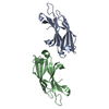
| Entry | Database: PDB / ID: 3q5m | ||||||
|---|---|---|---|---|---|---|---|
| Title | Crystal structure of Escherichia coli BamD | ||||||
 Components Components | UPF0169 lipoprotein yfiO | ||||||
 Keywords Keywords |  PROTEIN BINDING / Lipoprotein / BamD PROTEIN BINDING / Lipoprotein / BamD | ||||||
| Function / homology |  Function and homology information Function and homology informationBam protein complex / Gram-negative-bacterium-type cell outer membrane assembly / protein insertion into membrane / cell outer membrane / membraneSimilarity search - Function | ||||||
| Biological species |  Escherichia coli (E. coli) Escherichia coli (E. coli) | ||||||
| Method | X-RAY DIFFRACTION / SYNCHROTRON / SAD / Resolution: 2.604 Å | ||||||
 Authors Authors | Dong, C. / Hou, H. / Yang, X. / Dong, Y. / Shen, Y. | ||||||
 Citation Citation | Journal: Acta Crystallogr.,Sect.D / Year: 2012 Title: Structure of Escherichia coli BamD and its functional implications in outer membrane protein assembly Authors: Dong, C. / Hou, H.F. / Yang, X. / Shen, Y.Q. / Dong, Y.H. | ||||||
| History |
|
- Structure visualization
| Structure viewer | Molecule: MolmilJmol/JSmol |
|---|
- Downloads & links
-Download
| PDBx/mmCIF format | 3q5m.cif.gz | 97.5 KB | Display | PDBx/mmCIF format |
|---|---|---|---|---|
| PDB format | pdb3q5m.ent.gz | 76.1 KB | Display | PDB format |
| PDBx/mmJSON format | 3q5m.json.gz | Tree view | PDBx/mmJSON format | |
| Others |  Other downloads Other downloads |
-Validation report
| Arichive directory | https://data.pdbj.org/pub/pdb/validation_reports/q5/3q5mftp://data.pdbj.org/pub/pdb/validation_reports/q5/3q5m | HTTPS FTP |
|---|
-Related structure data
| Similar structure data |
|---|


-Links
 PDBj
PDBj


- Assembly
| Deposited unit | 
| ||||||||
|---|---|---|---|---|---|---|---|---|---|
| 1 |
| ||||||||
| 2 | 
| ||||||||
| Unit cell |
| ||||||||
| Components on special symmetry positions |
|
-Components
| #1: Protein | Mass: 25640.625 Da / Num. of mol.: 1 Source method: isolated from a genetically manipulated source Source: (gene. exp.) Escherichia coli (E. coli) / Strain: K12 / Gene: yfiO / Production host: Escherichia coli (E. coli) / References: UniProt: P0AC02 | ||
|---|---|---|---|
| #2: Chemical | ChemComp-IOD / Iodide  Mass: 126.904 Da / Num. of mol.: 7 / Source method: obtained synthetically / Formula: I Mass: 126.904 Da / Num. of mol.: 7 / Source method: obtained synthetically / Formula: I#3: Water | ChemComp-HOH / | Water Mass: 18.015 Da / Num. of mol.: 21 / Source method: isolated from a natural source / Formula: H2O Mass: 18.015 Da / Num. of mol.: 21 / Source method: isolated from a natural source / Formula: H2O |
-Experimental details
-Experiment
| Experiment | Method: X-RAY DIFFRACTION / Number of used crystals: 1 |
|---|
- Sample preparation
| Crystal | Density Matthews: 3.6 Å3/Da / Density % sol: 65.85 % |
|---|---|
| Crystal grow | Temperature: 277 K / Method: vapor diffusion, sitting drop / pH: 7 Details: 0.14M KI, 26%-27% PEG3350, pH 7.0, VAPOR DIFFUSION, SITTING DROP, temperature 277K |
-Data collection
| Diffraction | Mean temperature: 100 K |
|---|---|
| Diffraction source | Source: SYNCHROTRON / Site: BSRF  / Beamline: 3W1A / Wavelength: 0.9791 Å / Beamline: 3W1A / Wavelength: 0.9791 Å |
| Detector | Type: MARMOSAIC 325 mm CCD / Detector: CCD / Date: Jul 21, 2010 |
| Radiation | Monochromator: GRAPHITE / Protocol: SINGLE WAVELENGTH / Monochromatic (M) / Laue (L): M / Scattering type: x-ray |
| Radiation wavelength | Wavelength: 0.9791 Å / Relative weight: 1 |
| Reflection | Resolution: 2.6→50 Å / Num. all: 11964 / Num. obs: 10307 / % possible obs: 86.2 % / Observed criterion σ(F): 5 / Observed criterion σ(I): 2 / Redundancy: 23.3 % / Rmerge(I) obs: 0.107 / Rsym value: 0.133 / Net I/σ(I): 41.38 |
| Reflection shell | Resolution: 2.6→2.69 Å / Redundancy: 10.4 % / Rmerge(I) obs: 0.624 / Mean I/σ(I) obs: 2.8 / Num. unique all: 1155 / Rsym value: 0.643 / % possible all: 99.9 |
- Processing
| Software | Name: PHENIX / Version: (phenix.refine: 1.6.4_486) / Classification: refinement | |||||||||||||||||||||||||||||||||||||||||||||||||||||||||||||||
|---|---|---|---|---|---|---|---|---|---|---|---|---|---|---|---|---|---|---|---|---|---|---|---|---|---|---|---|---|---|---|---|---|---|---|---|---|---|---|---|---|---|---|---|---|---|---|---|---|---|---|---|---|---|---|---|---|---|---|---|---|---|---|---|---|
| Refinement | Method to determine structure: SAD / Resolution: 2.604→32.602 Å / SU ML: 0.44 / Cross valid method: THROUGHOUT / σ(F): 2.49 / σ(I): 2 / Stereochemistry target values: MLHL
| |||||||||||||||||||||||||||||||||||||||||||||||||||||||||||||||
| Solvent computation | Shrinkage radii: 0.95 Å / VDW probe radii: 1.2 Å / Solvent model: FLAT BULK SOLVENT MODEL / Bsol: 39.06 Å2 / ksol: 0.333 e/Å3 | |||||||||||||||||||||||||||||||||||||||||||||||||||||||||||||||
| Displacement parameters |
| |||||||||||||||||||||||||||||||||||||||||||||||||||||||||||||||
| Refinement step | Cycle: LAST / Resolution: 2.604→32.602 Å
| |||||||||||||||||||||||||||||||||||||||||||||||||||||||||||||||
| Refine LS restraints |
| |||||||||||||||||||||||||||||||||||||||||||||||||||||||||||||||
| LS refinement shell |
| |||||||||||||||||||||||||||||||||||||||||||||||||||||||||||||||
| Refinement TLS params. | Method: refined / Origin x: 37.665 Å / Origin y: -0.7724 Å / Origin z: 36.543 Å
| |||||||||||||||||||||||||||||||||||||||||||||||||||||||||||||||
| Refinement TLS group | Selection details: all |
