 Movie
Movie Controller

[English] 日本語
 Yorodumi
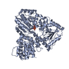
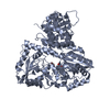
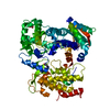
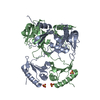
- PDB-3q3e: Crystal structure of the Actinobacillus pleuropneumoniae HMW1C gl... -
+ Open data

- Basic information
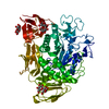
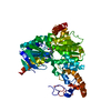
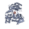
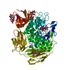
| Entry | Database: PDB / ID: 3q3e | ||||||
|---|---|---|---|---|---|---|---|
| Title | Crystal structure of the Actinobacillus pleuropneumoniae HMW1C glycosyltransferase | ||||||
 Components Components | HMW1C-like glycosyltransferase | ||||||
 Keywords Keywords |  TRANSFERASE / HMW1C / HMW1 / N-glycosylation TRANSFERASE / HMW1C / HMW1 / N-glycosylation | ||||||
| Function / homology | Rossmann fold - #11380 / Glycogen Phosphorylase B; / Rossmann fold / 3-Layer(aba) Sandwich / Alpha Beta / :  Function and homology information Function and homology information | ||||||
| Biological species |  Actinobacillus pleuropneumoniae serovar 1 (bacteria) Actinobacillus pleuropneumoniae serovar 1 (bacteria) | ||||||
| Method | X-RAY DIFFRACTION / SYNCHROTRON / SIR / Resolution: 2.1 Å | ||||||
 Authors Authors | Kawai, F. / Yeo, H.J. | ||||||
 Citation Citation | Journal: J.Biol.Chem. / Year: 2011 Title: Structural Insights into the Glycosyltransferase Activity of the Actinobacillus pleuropneumoniae HMW1C-like Protein. Authors: Kawai, F. / Grass, S. / Kim, Y. / Choi, K.J. / St Geme, J.W. / Yeo, H.J. | ||||||
| History |
|
- Structure visualization
| Structure viewer | Molecule: MolmilJmol/JSmol |
|---|
- Downloads & links
-Download
| PDBx/mmCIF format | 3q3e.cif.gz | 256.4 KB | Display | PDBx/mmCIF format |
|---|---|---|---|---|
| PDB format | pdb3q3e.ent.gz | 206.5 KB | Display | PDB format |
| PDBx/mmJSON format | 3q3e.json.gz | Tree view | PDBx/mmJSON format | |
| Others |  Other downloads Other downloads |
-Validation report
| Arichive directory | https://data.pdbj.org/pub/pdb/validation_reports/q3/3q3eftp://data.pdbj.org/pub/pdb/validation_reports/q3/3q3e | HTTPS FTP |
|---|
-Related structure data


-Links
 PDBj
PDBj- Assembly
| Deposited unit | 
| ||||||||
|---|---|---|---|---|---|---|---|---|---|
| 1 | 
| ||||||||
| 2 | 
| ||||||||
| Unit cell |
|
-Components
| #1: Protein | Mass: 71870.711 Da / Num. of mol.: 2 Source method: isolated from a genetically manipulated source Source: (gene. exp.) Actinobacillus pleuropneumoniae serovar 1 (bacteria)Strain: 4074 / Gene: APL_1635, appser1_17560 / Plasmid: PET45B / Production host: Escherichia coli (E. coli) / Strain (production host): BL21(DE3) / References: UniProt: E0EAD4#2: Chemical | Glycerol  Mass: 92.094 Da / Num. of mol.: 3 / Source method: obtained synthetically / Formula: C3H8O3 Mass: 92.094 Da / Num. of mol.: 3 / Source method: obtained synthetically / Formula: C3H8O3#3: Water | ChemComp-HOH / | Water Mass: 18.015 Da / Num. of mol.: 484 / Source method: isolated from a natural source / Formula: H2O Mass: 18.015 Da / Num. of mol.: 484 / Source method: isolated from a natural source / Formula: H2OSequence details | S428P POSSIBLE NATURAL VARIANT | |
|---|
-Experimental details
-Experiment
| Experiment | Method: X-RAY DIFFRACTION / Number of used crystals: 1 |
|---|
- Sample preparation
| Crystal | Density Matthews: 2.34 Å3/Da / Density % sol: 47.46 % |
|---|---|
| Crystal grow | Temperature: 288 K / Method: vapor diffusion, hanging drop / pH: 6.5 Details: 0.1 M MES, 0.1-0.16 M ammonium sulfate, 20-30% w/v PEG8000, pH 6.5, VAPOR DIFFUSION, HANGING DROP, temperature 288K |
-Data collection
| Diffraction | Mean temperature: 100 K |
|---|---|
| Diffraction source | Source: SYNCHROTRON / Site: APS  / Beamline: 19-ID / Wavelength: 0.97943 / Beamline: 19-ID / Wavelength: 0.97943 |
| Detector | Type: ADSC QUANTUM 315 / Detector: CCD / Date: Jul 16, 2009 |
| Radiation | Monochromator: SI 111 / Protocol: SINGLE WAVELENGTH / Monochromatic (M) / Laue (L): M / Scattering type: x-ray |
| Radiation wavelength | Wavelength: 0.97943 Å / Relative weight: 1 |
| Reflection | Resolution: 2.1→50 Å / Num. obs: 73875 / % possible obs: 93.3 % / Redundancy: 3.7 % / Rmerge(I) obs: 0.08 |
| Reflection shell | Resolution: 2.1→2.18 Å / Redundancy: 2.4 % / Rmerge(I) obs: 0.291 / % possible all: 83.3 |
- Processing
| Software |
| ||||||||||||||||||||||||||||||||||||||||||||||||||||||||||||||||||||||||||||||||||||||||||||||||||||||||||||||||||||||||||||||||||||||||||||||||||||||||||||||||||||||||||
|---|---|---|---|---|---|---|---|---|---|---|---|---|---|---|---|---|---|---|---|---|---|---|---|---|---|---|---|---|---|---|---|---|---|---|---|---|---|---|---|---|---|---|---|---|---|---|---|---|---|---|---|---|---|---|---|---|---|---|---|---|---|---|---|---|---|---|---|---|---|---|---|---|---|---|---|---|---|---|---|---|---|---|---|---|---|---|---|---|---|---|---|---|---|---|---|---|---|---|---|---|---|---|---|---|---|---|---|---|---|---|---|---|---|---|---|---|---|---|---|---|---|---|---|---|---|---|---|---|---|---|---|---|---|---|---|---|---|---|---|---|---|---|---|---|---|---|---|---|---|---|---|---|---|---|---|---|---|---|---|---|---|---|---|---|---|---|---|---|---|---|---|
| Refinement | Method to determine structure: SIR / Resolution: 2.1→50 Å / Cor.coef. Fo:Fc: 0.946 / Cor.coef. Fo:Fc free: 0.923 / SU B: 5.2 / SU ML: 0.133 / Cross valid method: THROUGHOUT / ESU R Free: 0.185 / Stereochemistry target values: MAXIMUM LIKELIHOOD
| ||||||||||||||||||||||||||||||||||||||||||||||||||||||||||||||||||||||||||||||||||||||||||||||||||||||||||||||||||||||||||||||||||||||||||||||||||||||||||||||||||||||||||
| Solvent computation | Ion probe radii: 0.8 Å / Shrinkage radii: 0.8 Å / VDW probe radii: 1.4 Å / Solvent model: BABINET MODEL WITH MASK | ||||||||||||||||||||||||||||||||||||||||||||||||||||||||||||||||||||||||||||||||||||||||||||||||||||||||||||||||||||||||||||||||||||||||||||||||||||||||||||||||||||||||||
| Displacement parameters | Biso mean: 25.41 Å2
| ||||||||||||||||||||||||||||||||||||||||||||||||||||||||||||||||||||||||||||||||||||||||||||||||||||||||||||||||||||||||||||||||||||||||||||||||||||||||||||||||||||||||||
| Refinement step | Cycle: LAST / Resolution: 2.1→50 Å
| ||||||||||||||||||||||||||||||||||||||||||||||||||||||||||||||||||||||||||||||||||||||||||||||||||||||||||||||||||||||||||||||||||||||||||||||||||||||||||||||||||||||||||
| Refine LS restraints |
| ||||||||||||||||||||||||||||||||||||||||||||||||||||||||||||||||||||||||||||||||||||||||||||||||||||||||||||||||||||||||||||||||||||||||||||||||||||||||||||||||||||||||||
| LS refinement shell | Resolution: 2.1→2.16 Å / Total num. of bins used: 20
|
